Category:Works of Raymond Sabouraud
Jump to navigation
Jump to search
Media in category "Works of Raymond Sabouraud"
The following 39 files are in this category, out of 39 total.
-
 Aspergillus niger Micrograph.jpg 640 × 480; 46 KB
-
 Candida spp.jpg 650 × 650; 84 KB
-
 Chrysosporium keratinophilum.jpg 697 × 697; 349 KB
-
 Coccidioides immitis on Sabouraud's medium.jpg 363 × 444; 44 KB
-
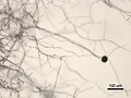
 Fungo dermatófito.jpg 2,048 × 1,828; 417 KB
-
 Geomyces destructans cultures.jpg 1,387 × 872; 747 KB
-
 Madura foot me photo for help in diagnosis.jpg 800 × 672; 367 KB
-
 Microsporum gypseum-003.JPG 1,185 × 1,185; 1.76 MB
-
 Mucor racemosus.jpeg 4,608 × 3,456; 3.62 MB
-
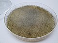
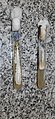
 Mycetoma culture.png 1,765 × 1,599; 3.99 MB
-
 Nectar Microbes - Corrugated.jpg 5,398 × 3,884; 12.54 MB
-
 Nectar Microbes - Guttation 2.jpg 5,322 × 3,443; 11.33 MB
-
 Nectar Microbes - Guttation.jpg 5,412 × 3,651; 11.49 MB
-
 Nectar Microbes - Lobate.jpg 5,199 × 3,732; 16.2 MB
-
 Nectar Microbes - Undulate.jpg 6,000 × 4,000; 23.04 MB
-

-
 Phaeoacremonium tardicrescens.png 2,615 × 2,615; 9.72 MB
-
 Radiography, x-ray therapeutics and radium therapy (1915) (14570898109).jpg 668 × 1,584; 139 KB
-
 Radiography, x-ray therapeutics and radium therapy (1915) (14571795307).jpg 1,802 × 2,868; 721 KB
-
 Radiography, X-ray therapeutics and radium therapy (1916) (14756014104).jpg 1,828 × 2,898; 548 KB
-
 Rhodotorula mucilaginosa colonies 45.jpg 1,200 × 1,200; 486 KB
-
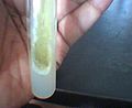
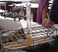 Sabouraud's glucose agar in a test tube.JPG 483 × 431; 43 KB
-
 Sabouraud's glucose agar.JPG 352 × 288; 41 KB
-
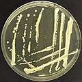
 Saccharomyces cerevisiae SAB colonies 33.jpg 900 × 900; 239 KB
-
 Saccharomyces cerevisiae SAB colonies 35.jpg 900 × 900; 266 KB
-
 T tonsurans colony 800.jpg 800 × 1,100; 719 KB
-
 The American journal of tropical medicine (1921) (14597746548).jpg 1,304 × 1,944; 691 KB
-
 The British journal of dermatology (1888) (14578895908).jpg 1,392 × 1,398; 442 KB
-
 The British journal of dermatology (1888) (14580493020).jpg 1,806 × 692; 79 KB
-
 The British journal of dermatology (1888) (14767197045).jpg 1,838 × 1,050; 193 KB
-
 The British journal of dermatology (1888) (14777977151).jpg 1,214 × 1,592; 441 KB
-
 Trichophyton rubrum colonies 800.jpg 800 × 800; 664 KB
-
 Trichophyton rubrum var. rodhaini PHIL 4248 lores.jpg 700 × 504; 45 KB
-
 Trichophyton terrestre PHIL 4300 lores.jpg 700 × 486; 41 KB
-
 Ttonsurans011.jpg 329 × 329; 120 KB
-

-
 Yeast and mold colony morphology on Sabouraud's dextrose agar (SDA).jpg 1,844 × 4,000; 2.77 MB
-
 Yeast cells in Gram staining of Candida growth on SDA.jpg 4,160 × 2,340; 1.46 MB
-
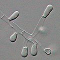
 Zygosaccharomyces bailii cells.jpg 690 × 516; 135 KB


_(14570898109).jpg)
_(14571795307).jpg)
_(14756014104).jpg)


_(14597746548).jpg)
_(14578895908).jpg)
_(14767197045).jpg)
_(14777977151).jpg)


_(14753796501).jpg)
.jpg)


_(14580493020).jpg)