Category:Wellcome Library medical illustrations
Jump to navigation
Jump to search
Subcategories
This category has the following 3 subcategories, out of 3 total.
Media in category "Wellcome Library medical illustrations"
The following 92 files are in this category, out of 92 total.
-
 "Ex typ. sub signo Stellae", 1662 Wellcome L0009792.jpg 1,190 × 1,652; 833 KB
-
 'De Motu Cordis', by William Harvey, Frankfurt, Germany, 162 Wellcome L0060483.jpg 4,256 × 2,832; 1.48 MB
-
 'Knee Jerk' from W R Gowers' Manual of Diseases of the Nervous System 1886.jpg 2,547 × 1,837; 1.42 MB
-
 'Part of the trunk of a crooked skeleton' Wellcome L0022317.jpg 1,246 × 1,722; 784 KB
-
 'Two views of the trunk of a crooked skeleton', 1733. Wellcome L0022318.jpg 1,184 × 1,702; 848 KB
-
 A double sheet showing various ophthalmology instruments, ey Wellcome V0016255.jpg 3,128 × 2,596; 3.96 MB
-
 A man holding his nose to avoid breathing in a miasma. Drawi Wellcome L0027123.jpg 4,258 × 5,335; 5.25 MB
-
 A sheet of eye examinations and diagrams of the eye with a n Wellcome V0015920.jpg 3,450 × 2,072; 4.28 MB
-
 A sheet of three diagrams showing inflamed Wellcome V0015922.jpg 648 × 486; 65 KB
-
 A sheet of three diagrams showing inflamed Wellcome V0015923.jpg 648 × 486; 67 KB
-
 A sheet showing optical instruments, eye examinations and an Wellcome V0015917.jpg 3,352 × 2,012; 3.75 MB
-
 A sheet showing optical instruments, eye examinations, and d Wellcome V0015921.jpg 3,359 × 2,020; 3.24 MB
-
 A sheet showing optical instruments, eye examinations, diagr Wellcome V0015918.jpg 3,425 × 2,047; 3.5 MB
-
 A sheet showing optical instruments, eye examinations, diagr Wellcome V0015919.jpg 3,332 × 2,064; 3.45 MB
-
 A. von Haller, Opuscula sua anatomica... Wellcome L0023727.jpg 1,100 × 1,660; 679 KB
-
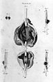
 A. von Haller, siamese twins. Wellcome L0021379.jpg 1,198 × 1,578; 902 KB
-
 A. von Haller, siamese twins. Wellcome L0021380.jpg 1,101 × 1,675; 1.09 MB
-
 A. Willett & W. Walsham "congenital malformation", plates Wellcome L0015301.jpg 1,146 × 1,838; 746 KB
-
 A. Willett & W. Walsham "congenital malformation", plates Wellcome L0015302.jpg 1,118 × 1,772; 807 KB
-
 A.C. Bock, Plate from Chirurgisch-Anatomische Tefeln... Wellcome L0003642.jpg 1,138 × 1,604; 843 KB
-
 Accidents caused by the use of green arsenic, 1859 Wellcome L0075299.jpg 6,471 × 4,745; 8.23 MB
-
 Accidents caused by the use of green arsenic, 1859 Wellcome L0075300.jpg 2,785 × 4,653; 4.52 MB
-
 Alexander Monro I; "Traite d'osteologie", 1759 Wellcome L0013268.jpg 1,100 × 1,714; 746 KB
-
 Anatomy of the ear, Thomas Buchanan, 1823 Wellcome L0035291.jpg 2,754 × 3,804; 2.47 MB
-
 Anatomy of the ear, Thomas Buchanan, 1823 Wellcome L0035292.jpg 3,156 × 3,845; 2.68 MB
-
 Apparatus for the treatment of paralysis Wellcome M0011330.jpg 3,763 × 2,881; 3.59 MB
-
 Arteries, 1801 Wellcome L0010509.jpg 1,144 × 1,692; 761 KB
-
 Arteries, 1801 Wellcome L0010510.jpg 1,688 × 1,112; 533 KB
-
 Back of human figure, spine; Vesling "Syntagma", 1647 Wellcome L0007897.jpg 1,174 × 1,622; 816 KB
-
 Bertillon, Instructions Signaletiques Album Wellcome L0030628.jpg 5,348 × 2,084; 2.73 MB
-
 Bertillon, Instructions Signaletiques Wellcome L0030629.jpg 1,106 × 1,778; 958 KB
-
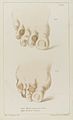
 Bones of the pelvis; two figures showing Wellcome V0008822.jpg 2,406 × 3,568; 3.63 MB
-
 Bourgery & Jacob, "Traite complet de l'anatomie de l'homme" Wellcome L0014791.jpg 1,172 × 1,628; 714 KB
-
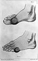 Bunions Wellcome L0006822.jpg 1,106 × 1,754; 884 KB
-
 C. Bell, A series of engravings.... Wellcome L0009980.jpg 1,036 × 1,812; 458 KB
-
 Charles Bell, Engravings of the Arteries Wellcome L0022057.jpg 1,124 × 1,748; 503 KB
-
 Depictions of the sexual organs of the male, the female and Wellcome V0038933.jpg 2,544 × 2,946; 4.01 MB
-
 Diagram showing incisions performed in caesarean operation Wellcome L0038227.jpg 2,706 × 3,936; 3.31 MB
-
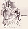 Diagrammatic View of the Ear.jpg 1,033 × 1,088; 1.33 MB
-
 Diagrams of incisors, canines and molars, a diseased tooth a Wellcome V0011997.jpg 2,126 × 3,530; 3.77 MB
-
 Dieffenbach, Chirurgische Erfahrungen Wellcome L0028279.jpg 1,200 × 1,646; 872 KB
-
 Distempered skin, John Machin Wellcome L0009888.jpg 1,490 × 1,242; 804 KB
-
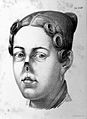
 Down syndrome lg.jpg 575 × 633; 28 KB
-
 Drawings of pathological subjects by B.E. Nicholson, 1946-50 Wellcome L0019366.jpg 1,274 × 1,502; 1.06 MB
-
 EB1911 Intestinal Obstruction - strangulation by a band.jpg 267 × 250; 31 KB
-
 Encephalitis, in Specilegium anatomicum Wellcome L0005443.jpg 1,162 × 1,580; 827 KB
-
 Engraving showing the diseased part of a skull Wellcome L0034202.jpg 3,796 × 3,076; 5.44 MB
-
 Engravings illustrating diseases of arteries. Wellcome L0011706.jpg 1,888 × 1,064; 858 KB
-
 Eye diseases and surgical instruments. Line engraving by F. Wellcome V0015915.jpg 3,348 × 2,060; 3.45 MB
-
 Feet from La Forest, L'art de soigner les pieds. Wellcome L0013757.jpg 5,800 × 3,166; 5.92 MB
-
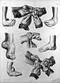
 Feet showing different types of bunions Wellcome L0039107.jpg 2,496 × 3,936; 4.15 MB
-
 Feet showing different types of corns Wellcome L0039106.jpg 2,400 × 3,936; 3.51 MB
-
 Forearm wound. Pooll. Captain of the Wassenaer. Wellcome L0033438.jpg 3,032 × 4,152; 2.42 MB
-
 Gautier D'Agoty; Anatomie de la tete... Wellcome L0023747.jpg 1,561 × 1,157; 705 KB
-
 Illustration deformed female pelvis - elliptical distortion Wellcome L0038230.jpg 2,520 × 3,996; 2.79 MB
-
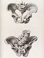 Illustration of a deformed female pelvis - angular distortion Wellcome L0038229.jpg 2,628 × 3,465; 2.58 MB
-
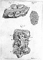
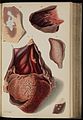
 Illustrations of a diseased heart, 1834 Wellcome L0074291.jpg 5,484 × 7,912; 12.09 MB
-
 Illustrations of a diseased heart, 1834 Wellcome L0074292.jpg 5,436 × 7,888; 15.48 MB
-
 Insertion of syphon trocar into ovarian cyst Wellcome L0014479.jpg 1,690 × 1,144; 593 KB
-
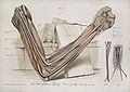
 J. Parsons, A mechanical and critical enquiry Wellcome L0022324.jpg 3,978 × 5,508; 6.19 MB
-
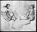
 J. von Heine, Poliomyelitis, 19th C. Wellcome L0003129.jpg 1,240 × 1,526; 638 KB
-
 J. von Heine, Poliomylitis, 19thC. Wellcome L0003130.jpg 1,236 × 1,562; 704 KB
-
 Lister spray in use from Cheyne, Antiseptic Surgery. Wellcome L0004098.jpg 3,760 × 2,825; 3.85 MB
-
 Medico-chirurgical transactions, volume 27 Wellcome L0030131.jpg 1,569 × 1,346; 1.02 MB
-
 Muscles of the back in a female - Wellcome Library, London - L0019727.jpg 3,638 × 4,835; 8.57 MB
-
 Operations for club-foot. Wellcome L0014790.jpg 1,164 × 1,624; 702 KB
-
 Pathological Anatomy; three sections of an aneurysm Wellcome L0019370.jpg 1,479 × 1,270; 887 KB
-
 Photo of obese man with Infantilism and degenerative disorder Wellcome L0034947.jpg 3,714 × 4,962; 6.67 MB
-
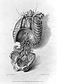
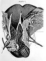 R. Lee, Ganglionic nervous plexuses... Wellcome L0004303.jpg 1,204 × 1,594; 789 KB
-
 Scarpa "Aneurism", 1819; plate Wellcome L0004113.jpg 1,094 × 1,678; 670 KB
-
 Superficial blood vessels of the head and neck.jpg 956 × 640; 195 KB
-
 Surgical Anatomy of the Arteries of the neck Gray's Anatomy 1858.jpg 470 × 548; 114 KB
-
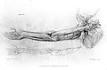
 The effect of Leprosey on the arm nerve Henry Vandyke Carter.jpg 780 × 550; 108 KB
-
 Three diagrams illustrating how to bleed an arm. Stipple eng Wellcome L0005148.jpg 1,206 × 1,680; 957 KB
-
 Treatment of hemiplegia Wellcome M0011329.jpg 2,921 × 3,732; 2.28 MB
-
 A vaccine pustule on the 7th to 8th day. Watercolour, c. 180 Wellcome V0016657.jpg 2,473 × 2,894; 2.82 MB
-
 A vaccine pustule on the 8th to 9th day. Watercolour, c. 180 Wellcome V0016658.jpg 2,668 × 3,168; 3.01 MB
-
 A comparison between a cowpox and smallpox pustule on the ei Wellcome V0016666.jpg 2,952 × 2,385; 2.41 MB
-
 A smallpox pustule on the eighth day of the disease. Waterco Wellcome V0016667.jpg 2,470 × 2,982; 2.4 MB
-
 Cowpox pustules from the sixth to the eighteenth days of the Wellcome V0016668.jpg 3,066 × 3,624; 1.86 MB
-
 A comparison between smallpox and cowpox pustules on the 8th Wellcome V0016672.jpg 2,250 × 3,120; 3.23 MB
-
 A comparison between smallpox and cowpox pustules on the 10t Wellcome V0016673.jpg 2,300 × 3,121; 3.29 MB
-
 A cow's udder with vaccinia pustules. Chromolithograph. Wellcome V0016679.jpg 2,270 × 3,114; 2.97 MB
-
 A doctor administers a clyster to a greedy little boy who is Wellcome V0016685.jpg 2,322 × 3,203; 3.39 MB
-
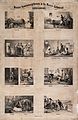 Hydrotherapy; eight vignettes of different cures at Wellcome V0016688.jpg 2,174 × 3,340; 3.58 MB
-
 Sixteen diagrams illustrating different bandages and how to Wellcome V0016820.jpg 2,419 × 3,182; 3.6 MB
-
 Diagrams illustrating how to bandage and set fractures in sp Wellcome V0016830.jpg 2,066 × 3,442; 3.47 MB
-
 A man sitting in a chair reading whilst his leg is in tracti Wellcome V0016833.jpg 2,420 × 2,896; 2.75 MB
-
 Two human figures illustrating the reduction of dislocations Wellcome V0016835.jpg 2,987 × 2,812; 3.5 MB
-
 A country barber shaving a farmer; another farmer looks into Wellcome V0016975.jpg 2,406 × 3,070; 3.16 MB
-
 Varicocele from La'mert, Self-preservation... Wellcome L0017942.jpg 1,080 × 1,752; 885 KB
-
 W. Cheselden, 1750; hermaphrodites Wellcome L0018091.jpg 1,073 × 1,766; 915 KB


