Category:Transmission electron microscopic images of Mus musculus
Jump to navigation
Jump to search
Media in category "Transmission electron microscopic images of Mus musculus"
The following 9 files are in this category, out of 9 total.
-
Bronchiolar area cilia cross-sections 1.jpg 1,600 × 1,278; 776 KB
-
Junctional complex and pinocytotic vesicles - embryonic brain-TEM.jpg 1,600 × 1,278; 939 KB
-
Lung epithelium 80294-2.6.jpg 1,600 × 1,286; 829 KB
-
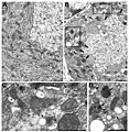
Mitophagy purkinje cell.jpg 600 × 617; 142 KB
-
Orientia tsutsugamushi.JPG 2,848 × 2,224; 1.39 MB
-
Sin Nombre hanta virus TEM PHIL 1136 lores.jpg 3,060 × 2,033; 1.97 MB
-
Spindle centriole - embryonic brain mouse - TEM.jpg 1,283 × 1,600; 901 KB
-
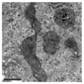
TEM image of mitochondria in MEF OMA1-KO after PXA.png 490 × 490; 170 KB
-
Unctional complex and pinocytotic vesicles - embryonic brain - TEM.jpg 1,600 × 1,278; 861 KB


