Category:Toxoplasma
Jump to navigation
Jump to search
Domain: Eukaryota • Regnum: Protista • Superphylum: Alveolata • Phylum: Apicomplexa • Classis: Conoidasida • Subclassis: Coccidiasina • Ordo: Eucoccidiorida • Familia: Sarcocystidae • Genus: Toxoplasma

Wikispecies has an entry on:
obligate intracellular parasitic protozoan that causes toxoplasmosis | |||||||||||||||||||||||||||||
| Upload media | |||||||||||||||||||||||||||||
| Instance of | |||||||||||||||||||||||||||||
|---|---|---|---|---|---|---|---|---|---|---|---|---|---|---|---|---|---|---|---|---|---|---|---|---|---|---|---|---|---|
| |||||||||||||||||||||||||||||
| |||||||||||||||||||||||||||||
| Original combination | Leishmania gondii | ||||||||||||||||||||||||||||
| Taxon author | Charles Nicolle, 1908 | ||||||||||||||||||||||||||||
| |||||||||||||||||||||||||||||
Subcategories
This category has the following 3 subcategories, out of 3 total.
Media in category "Toxoplasma"
The following 48 files are in this category, out of 48 total.
-
Active-toxoplasmic-retinochoroiditis.jpg 600 × 507; 90 KB
-
Apocryptovirus veae040f3.jpg 1,000 × 549; 113 KB
-
Apocryptovirus veae040f4.jpg 1,003 × 518; 144 KB
-
Apocryptovirus veae040f8.jpg 675 × 511; 86 KB
-
Cycle Toxoplasma gondii nltxt.jpg 733 × 720; 267 KB
-
IRG activation following pathogen entry.jpg 949 × 725; 156 KB
-
Parasite (journal) 2013, 20, 11 Costache - Toxoplasma.pdf 1,233 × 1,747, 5 pages; 556 KB
-
Parasite140105-fig3 Toxoplasmosis in a bar-shouldered dove - TEM of 2 tachyzoites.tif 1,378 × 1,683; 2.11 MB
-
Struc1-1.png 912 × 577; 137 KB
-
Struc1-2.png 912 × 577; 235 KB
-
Struc3.png 414 × 371; 91 KB
-
T gondii IFA.jpg 200 × 200; 2 KB
-
T gondii Taquizoito Conoide.png 387 × 757; 344 KB
-
T gondii Taquizoito.png 390 × 744; 291 KB
-
T-gondii Citoesqueleto.png 722 × 1,071; 433 KB
-
T-gondii Complejo-Apical.png 388 × 743; 297 KB
-
T-gondii Conoide microtu.png 1,266 × 632; 824 KB
-
T-gondii Estructura Conoide.png 1,170 × 2,077; 1.4 MB
-
T-gondii Estructura.png 2,790 × 1,283; 530 KB
-
T-gondii Intracelular.png 1,170 × 1,166; 1.7 MB
-
T-gondii Invasion.png 739 × 742; 205 KB
-
T-gondii.png 552 × 742; 264 KB
-
T. gondii tachyzoites.jpg 3,264 × 2,448; 608 KB
-
T. gondii traquizoito Conoide.png 1,200 × 1,035; 1.27 MB
-
Test d'invasion de fibroblastes par des parasites Toxoplasma gondii.jpg 2,625 × 2,006; 549 KB
-
ToRCH IgG Combo Rapid Test-Negative.jpg 3,264 × 2,448; 1.93 MB
-
ToRCH IgM and IgG Rapid Test-Negative.jpg 3,264 × 2,448; 1.86 MB
-
ToRCH IgM Combo Rapid Test-Negative.jpg 3,264 × 2,448; 1.89 MB
-
TORCH test device.jpg 6,000 × 8,000; 9.15 MB
-
ToxoCystUnstained.jpg 300 × 300; 15 KB
-
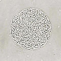
ToxoOocystFecalFlotation.jpg 300 × 300; 19 KB
-
Toxoplasma gondii (2).jpg 400 × 400; 91 KB
-
Toxoplasma gondii bradyzoite.png 917 × 971; 95 KB
-
Toxoplasma gondii gamete.png 1,204 × 789; 137 KB
-
Toxoplasma gondii merozoite.png 760 × 1,017; 60 KB
-
Toxoplasma gondii oocyst spore.png 1,513 × 1,613; 530 KB
-
Toxoplasma gondii oocyst.png 1,118 × 1,157; 188 KB
-
Toxoplasma gondii sporocyst.png 979 × 1,176; 288 KB
-
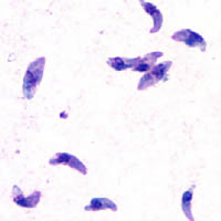
Toxoplasma gondii tachy.jpg 200 × 200; 5 KB
-
Toxoplasma gondii, Ausstrich.jpg 3,872 × 2,592; 2.08 MB
-
Toxoplasma gondii.jpg 2,625 × 2,625; 1.52 MB
-
TOXOPLASMA GONDII.jpg 3,896 × 2,617; 1.73 MB
-
Toxoplasma life cycle de.png 1,216 × 745; 398 KB
-
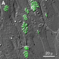
ToxoTachyzoitesGreen.jpg 300 × 300; 3 KB
-
Toxplasma.png 543 × 458; 172 KB


