Category:The Armed Forces Institute of Pathology Public Domain Images
Jump to navigation
Jump to search
The AFIP Electronic Fascicles are CD-ROM versions of the AFIP Atlas of Tumor Pathology. The Electronic Fascicles contain both U.S. Government work and copyrighted materials used with permission of non-Government contributors. Images that represent Government contributors are in public domain and have been included in the PEIR Digital Library, which is the source of images included in this category.
Media in category "The Armed Forces Institute of Pathology Public Domain Images"
The following 130 files are in this category, out of 130 total.
-
 Adamantinomatous craniopharyngioma.jpg 631 × 512; 210 KB
-
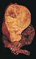
 Adenocarcinoma metastatic to adrenal gland.jpg 646 × 512; 220 KB
-
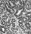
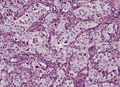
 Adenocarcinoma of the lung.jpg 703 × 512; 342 KB
-
 Adrenal cortical carcinoma.JPG 721 × 512; 292 KB
-
 Adrenal cortical carcinoma.jpg 720 × 512; 323 KB
-
 ALL, PAS.jpg 733 × 512; 259 KB
-
 ALL-L1.jpg 357 × 512; 144 KB
-
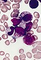
 AML-M0.jpg 355 × 512; 140 KB
-
 AML-M1.jpg 737 × 512; 288 KB
-
 AML-M2 associated with a t(8;21) chromosome abnormality.jpg 759 × 512; 304 KB
-
 AML-M2.jpg 351 × 512; 145 KB
-
 AML-M3.jpg 722 × 512; 300 KB
-
 AML-M3V.jpg 743 × 512; 279 KB
-
 AML-M4.jpg 726 × 512; 275 KB
-
 AML-M4EO.jpg 739 × 512; 256 KB
-
 AML-M5A.jpg 745 × 512; 332 KB
-
 AML-M5B, promonocyte, TEM.jpg 685 × 512; 309 KB
-
 AML-M5B.jpg 741 × 512; 292 KB
-
 AML-M6, blood smear(Right Rotation).png 417 × 442; 333 KB
-
 AML-M6, blood smear.jpg 737 × 512; 269 KB
-
 AML-M6, multinucleated erythroblast.jpg 725 × 512; 291 KB
-
 AML-M7, bone marrow section.jpg 359 × 512; 222 KB
-
 Atrial myxoma embolus.jpg 800 × 508; 279 KB
-
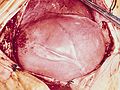
 Atrial myxoma.jpg 621 × 512; 270 KB
-
 AVM grossly.jpg 767 × 512; 254 KB
-
 AVM slice.jpg 800 × 507; 326 KB
-
 B-cell chronic lymphocytic leukemia (B-CLL).jpg 356 × 512; 159 KB
-
 Barrett's mucosa, Alcian blue stain.jpg 719 × 512; 374 KB
-
 Barrett's mucosa, H&E.jpg 734 × 512; 353 KB
-
 Barrett's mucosa, high-grade dysplasia, H&E.jpg 720 × 512; 372 KB
-

-
 Barrett's mucosa, high-grade dysplasia.jpg 364 × 512; 170 KB
-
 Barrett's mucosa, higher magnification, Alcian blue stain .jpg 721 × 512; 359 KB
-
 Barrett's mucosa, higher magnification, Alcian blue stain.jpg 719 × 512; 346 KB
-
 Barrett's mucosa, low-grade dysplasia, H&E.jpg 358 × 512; 181 KB
-
 Barrett's mucosa, low-grade dysplasia, high-power magnification.jpg 356 × 512; 170 KB
-
 Barrett's mucosa, low-grade dysplasia, low-power view, H&E.jpg 357 × 512; 201 KB
-
 Barrett's mucosa, low-grade dysplasia.jpg 735 × 512; 376 KB
-
 Barrett's mucosa, PAS-Alcian blue stain.jpg 364 × 512; 173 KB
-
 Beckwith.jpg 369 × 512; 197 KB
-
 Bilateral pheo MEN2.jpg 369 × 512; 151 KB
-
 Blood smear of anisopoikilocytosis in polycythemia vera.jpg 498 × 319; 38 KB
-
 Bronchioloalveolar carcinoma.jpg 738 × 512; 316 KB
-
 Carcinoid tumor with postobstructive pneumonia.jpg 703 × 512; 288 KB
-
 Cardiac rhabdomyoma .jpg 685 × 512; 299 KB
-
 Cardiac rhabdomyoma.jpg 683 × 512; 321 KB
-
 Central neurocytoma micro.jpg 724 × 512; 310 KB
-
 Clear cell adenocarcinoma of the lung.jpg 797 × 512; 382 KB
-
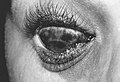
 Coats disease.jpg 492 × 512; 166 KB
-
 Diffuse pleural mesothelioma.jpg 719 × 512; 278 KB
-
 Diffuse pleural metastasis.jpg 434 × 512; 203 KB
-
 Dig micro1.jpg 468 × 512; 191 KB
-
 Encapsulated cystic thymoma.jpg 800 × 473; 286 KB
-
 Encapsulated thymoma.jpg 309 × 512; 117 KB
-
 Ependymoblastoma.jpg 472 × 512; 230 KB
-
 Ependymoma in the fourth ventricle.jpg 544 × 512; 197 KB
-
 Faggot cell in AML-M3.jpg 352 × 512; 145 KB
-
 Firbrous plaque on the diaphragm of an asbestos worker.jpg 721 × 512; 306 KB
-
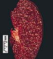 Follicular lymphoma, spleen.jpg 459 × 512; 179 KB
-
 Gastrinoma.jpg 729 × 512; 339 KB
-
 Glioblastoma multiforme.jpg 558 × 512; 180 KB
-
 Hamartoma of the lung.jpg 676 × 512; 281 KB
-
 Hippel angiogram.jpg 724 × 512; 240 KB
-
 Hodgkin's disease, nodular sclerosis.jpg 382 × 512; 125 KB
-
 Hodgkin's disease, spleen.jpg 677 × 512; 280 KB
-
 Hypogranular neutrophil with a pseudo-Pelger-Huet nucleus in MDS.jpg 359 × 512; 132 KB
-
 Hypothalamic hamartoma.jpg 619 × 512; 200 KB
-
 Islet hyperplasia BWS.jpg 771 × 512; 307 KB
-
 Keratoacanthoma.jpg 632 × 512; 233 KB
-
 Large cell carcinoma of the lung .jpg 701 × 512; 324 KB
-
 Large cell carcinoma of the lung.jpg 711 × 512; 308 KB
-
 Leptomeningeal fibrosarcoma.jpg 647 × 512; 215 KB
-
 Locally invasive circumscribed thymoma.jpg 311 × 512; 134 KB
-
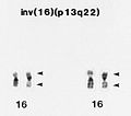 M4EO, pericentric inversion of chromosome 16.jpg 579 × 512; 157 KB
-
 Malignant melanoma.jpg 616 × 512; 223 KB
-
 Mature teratoma invading the lung.jpg 729 × 512; 279 KB
-
 Mature teratoma, mediastinum.jpg 595 × 512; 185 KB
-
 Mediastinal paraganglioma.jpg 614 × 512; 233 KB
-
 Medulloblastoma .jpg 800 × 476; 340 KB
-
 Medulloepithelioma.jpg 481 × 512; 171 KB
-
 Megakaryocytes in MDS (RAEB and 5q-chromosome abnormality).jpg 739 × 512; 252 KB
-
 Menetrier.jpg 721 × 512; 280 KB
-
 Meningioma.jpg 657 × 512; 216 KB
-
 Meta nasoph ca.jpg 459 × 512; 234 KB
-
 Metastatic carcinoma, brain.jpg 703 × 512; 227 KB
-
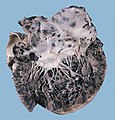 Metastatic malignant melanoma, heart.jpg 489 × 512; 216 KB
-
 MRI of a Dysembryoplastic Neuroepithelial Tumor.jpg 404 × 512; 119 KB
-
 Mucinous bronchioloalveolar carcinoma, digested PAS stain.jpg 704 × 512; 350 KB
-
 Myeloblast with Auer rod.jpg 164 × 133; 9 KB
-
 Myxoma.jpg 722 × 512; 258 KB
-
 Myxopapillary ependymoma 1.jpg 794 × 512; 300 KB
-
 Myxopapillary ependymoma.jpg 716 × 512; 321 KB
-
 Neuroblastoma in situ.jpg 711 × 512; 315 KB
-
 Neuroblastoma infant.jpg 347 × 512; 135 KB
-
 Neuroblastoma Muffin.jpg 724 × 512; 261 KB
-
 Neurocytaneous melanosis.jpg 435 × 512; 163 KB
-
 Neurothekeoma.jpg 248 × 512; 104 KB
-
 Nf2.jpg 637 × 512; 197 KB
-
 Nonmucinous bronchioloalveolar carcinoma.jpg 371 × 512; 182 KB
-
 Olfactory neuroblastoma.jpg 463 × 512; 199 KB
-
 Optic glioma.jpg 442 × 512; 141 KB
-
 Ota.jpg 746 × 512; 228 KB
-
 Papillary craniopharyngioma.jpg 596 × 512; 201 KB
-
 Persistent hyperplastic primary vitreous.jpg 455 × 512; 196 KB
-
 Philadelphia chromosome, t(9;22) translocation.jpg 655 × 512; 174 KB
-
 Pilocytic astrocytoma of the hypothalamic region.jpg 562 × 512; 190 KB
-
 Polycythemia vera, blood smear.jpg 5,872 × 4,096; 6.85 MB
-
 Pontine astrocytoma.jpg 710 × 512; 251 KB
-
 Poorly differentiated (solid) adenocarcinoma of the lung.jpg 707 × 512; 379 KB
-
 Psammoma.jpg 725 × 512; 323 KB
-
 Pulmonal multinodular metastasis of a renal cell carcinoma.jpg 699 × 512; 363 KB
-
 Radial scar.jpg 337 × 512; 150 KB
-
 Retinoblastoma .jpg 346 × 512; 123 KB
-
 Retinoblastoma rosette.jpg 368 × 512; 192 KB
-
 Rhabdoid tumor.jpg 725 × 512; 273 KB
-
 Rosai-dorfman.jpg 465 × 512; 218 KB
-
 Sega gross.jpg 717 × 512; 221 KB
-
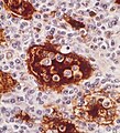
 Small cell carcinoma of the lung.jpg 704 × 512; 334 KB
-
 Spitz nevus.jpg 800 × 497; 269 KB
-
 Squamous cell carcinoma involving a subsegmental bronchus.jpg 711 × 512; 326 KB
-
 Subependymoma.jpg 800 × 484; 308 KB
-
 Tuberous sclerosis, cross section.jpg 789 × 512; 257 KB
-
 Tuberous sclerosis.jpg 561 × 512; 190 KB
-
 Tumor embolus in the main pulmonary artery.jpg 706 × 512; 329 KB
-

-
 Two myeloblasts with Auer rods.jpg 352 × 512; 123 KB
-
 Typical carcinoid tumor of the lung, prominent rosettes.jpg 404 × 512; 220 KB
-
 Typical carcinoid tumor of the lung, trabecular pattern.jpg 780 × 512; 403 KB
-
 Well differentiated squamous cell carcinoma.jpg 785 × 512; 391 KB
-
 Wilms tumor.jpg 776 × 512; 310 KB


_chromosome_abnormality.jpg)


.png)


.jpg)


.jpg)


_adenocarcinoma_of_the_lung.jpg)


_translocation.jpg)