Category:Taken with Olympus LC30
Jump to navigation
Jump to search
English: Taken with Olympus LC30 microscope cameras.
Media in category "Taken with Olympus LC30"
The following 104 files are in this category, out of 104 total.
-
Chromogenic immunohistochemistry for calponin in sclerosing adenosis.jpg 872 × 654; 173 KB
-
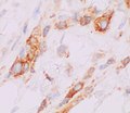
Chromogenic immunohistochemistry for E-cadherin in invasive lobular carcinoma.jpg 2,048 × 1,532; 271 KB
-
Chromogenic immunohistochemistry for p120 in invasive lobular carcinoma.jpg 1,549 × 1,347; 304 KB
-
Cross-section of whipworm on microscopy, original.jpg 983 × 613; 151 KB
-
Cross-section of whipworm on microscopy.jpg 849 × 773; 140 KB
-
Enterobius vermicularis - intermediate magnification.jpg 2,048 × 1,532; 329 KB
-
Enterobius vermicularis egg.jpg 603 × 417; 58 KB
-
Grade 2 clear cell renal cell carcinoma, original.jpg 2,048 × 1,532; 304 KB
-
Histology of anterior pituitary eosinophilic follicles.jpg 1,709 × 1,245; 543 KB
-
Histology of calcification of a term placenta.jpg 2,048 × 1,532; 495 KB
-
Histology of liver tissue from a gallbladder resection.jpg 2,048 × 1,532; 585 KB
-
Histology of macular artifacts in the hippocampus.jpg 2,048 × 1,532; 838 KB
-
Histology of paneth cells, annotated.jpg 1,257 × 1,117; 367 KB
-
Histology of paneth cells, original.jpg 2,048 × 1,532; 474 KB
-
Histology of thalamic neuron.jpg 2,048 × 1,532; 431 KB
-
Histomathology of meconium histocytosis (original).jpg 2,048 × 1,532; 448 KB
-
Histopatholgoy of acute gangrenous cholecystitis.jpg 2,048 × 1,532; 894 KB
-
Histopathology of a biopsy of a melanoma metastasis to the brain.jpg 2,048 × 1,532; 470 KB
-
Histopathology of a fresh thrombus, low magnification.jpg 2,048 × 1,532; 898 KB
-
Histopathology of a fundic glad polyp, low magnification.jpg 1,821 × 1,441; 592 KB
-
Histopathology of a hyperplastic polyp of the gallbladder.jpg 3,081 × 1,937; 1.43 MB
-
Histopathology of a keloid.jpg 2,048 × 1,532; 621 KB
-
Histopathology of acute choriodeciduitis (original).jpg 2,048 × 1,532; 483 KB
-
Histopathology of acute intraluminal inflammation of the appendix.jpg 2,048 × 1,532; 662 KB
-
Histopathology of amnionitis, annotated.jpg 1,195 × 771; 222 KB
-
Histopathology of amnionitis.jpg 2,048 × 1,532; 438 KB
-
Histopathology of avascular necrosis.jpg 2,048 × 1,532; 412 KB
-
Histopathology of bony tissue in a mature teratoma.jpg 2,048 × 1,532; 445 KB
-
Histopathology of brain tissue in a mature teratoma.jpg 2,048 × 1,532; 914 KB
-
Histopathology of cartwheel pattern in dermatofibrosarcoma protuberans, original.jpg 2,048 × 1,532; 603 KB
-
Histopathology of chondroid syringoma.jpg 2,048 × 1,532; 876 KB
-
Histopathology of chorioamnionitis.jpg 1,187 × 893; 254 KB
-
Histopathology of choroid plexus tissue in a mature teratoma.jpg 2,048 × 1,532; 800 KB
-
Histopathology of chronic pulmonary congestion.jpg 1,109 × 977; 173 KB
-
Histopathology of collagenous colitis.jpg 1,057 × 871; 232 KB
-
Histopathology of colorectal adenocarcinoma with lymphatic invasion.jpg 1,657 × 1,277; 640 KB
-
Histopathology of enterobius vermicularis eggs, HE stain.jpg 787 × 827; 125 KB
-
Histopathology of fatty tissue in a mature teratoma.jpg 2,048 × 1,532; 551 KB
-
Histopathology of flat epithelial atypia and columnar cell change.jpg 1,015 × 805; 244 KB
-
Histopathology of Gandy–Gamna nodules in chronic pulmonary congestion.jpg 2,048 × 1,532; 356 KB
-
Histopathology of glioblastoma, high magnification, annotated.jpg 1,453 × 997; 368 KB
-
Histopathology of glioblastoma, high magnification.jpg 2,048 × 1,532; 487 KB
-
Histopathology of goblet cells and foveolar cells in incomplete Barrett's esophagus.jpg 2,048 × 1,532; 665 KB
-
Histopathology of interstitial fibrosis of chronic ischemic heart disease.jpg 1,657 × 1,532; 821 KB
-
Histopathology of invasive melanoma, high magnification.jpg 2,048 × 1,532; 412 KB
-
Histopathology of inverted urothelial papilloma, high magnification.jpg 1,530 × 1,522; 640 KB
-
Histopathology of inverted urothelial papilloma.jpg 1,777 × 1,433; 835 KB
-
Histopathology of ischemic stroke of the thalamus at approximately 24 hours.jpg 2,048 × 1,532; 358 KB
-
Histopathology of lamina propria edema and hemorrhage in acute cholecystitis.jpg 2,048 × 1,532; 689 KB
-
Histopathology of leiomyoma with nuclear pleomorphism.jpg 2,048 × 1,532; 428 KB
-
Histopathology of lymphocytic colitis, annotated.jpg 2,048 × 1,532; 713 KB
-
Histopathology of lymphocytic colitis.jpg 2,048 × 1,532; 485 KB
-
Histopathology of lymphocytic myocarditis with myocyte necrosis, annotated.jpg 1,397 × 1,087; 385 KB
-
Histopathology of medullary breast carcinoma.jpg 2,048 × 1,532; 650 KB
-
Histopathology of mild active gastritis, annotated.jpg 2,048 × 1,532; 588 KB
-
Histopathology of mild active gastritis.jpg 2,048 × 1,532; 387 KB
-
Histopathology of mitral valve with myxomatous degeneration, annotated.jpg 2,397 × 1,417; 680 KB
-
Histopathology of mitral valve with myxomatous degeneration.jpg 2,397 × 1,417; 686 KB
-
Histopathology of moderate myocardial hypertrophy - boxcar nuclei.jpg 1,099 × 595; 182 KB
-
Histopathology of moderate myocardial hypertrophy.jpg 933 × 769; 190 KB
-
Histopathology of moderately differentiated esophageal adenocarcinoma.jpg 2,048 × 1,532; 509 KB
-
Histopathology of myocarditis with myocyte necrosis.jpg 2,048 × 1,532; 464 KB
-
Histopathology of neutrophil infiltration in myocardial infarction.jpg 1,804 × 1,218; 437 KB
-
Histopathology of osteomyelitis.jpg 1,497 × 1,067; 326 KB
-
Histopathology of pancreatic tissue in a mature cystic teratoma.jpg 2,048 × 1,532; 673 KB
-
Histopathology of papillary renal cell carcinoma type 1, high magnification.jpg 2,048 × 1,532; 521 KB
-
Histopathology of papillary renal cell carcinoma type 1, low magnification.jpg 2,048 × 1,532; 642 KB
-
Histopathology of parathyroid adenoma.jpg 2,048 × 1,532; 524 KB
-
Histopathology of paratubal cyst.jpg 453 × 381; 48 KB
-
Histopathology of phlebitis and funisitis, annotated.jpg 962 × 720; 224 KB
-
Histopathology of pleomorphic lobular carcinoma with plasmacytoid cells.jpg 2,048 × 1,532; 433 KB
-
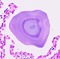
Histopathology of pulmonary alveolar microlithiasis.jpg 1,505 × 1,477; 341 KB
-
Histopathology of pyogenic granuloma - high magnification.jpg 2,048 × 1,532; 664 KB
-
Histopathology of pyogenic granuloma, high magnification, annotated.jpg 2,048 × 1,532; 682 KB
-
Histopathology of reactive inflammatory changes in a hyperplastic polyp.jpg 1,917 × 1,061; 702 KB
-
Histopathology of sclerosing adenosis of the breast.jpg 2,048 × 1,532; 493 KB
-
Histopathology of siderophage in chronic pulmonary congestion.jpg 883 × 833; 115 KB
-
Histopathology of squamous cell carcinoma of the urinary bladder, high magnification.jpg 2,048 × 1,532; 433 KB
-
Histopathology of squamous cell carcinoma of the urinary bladder, low magnification.jpg 2,048 × 1,532; 855 KB
-
Histopathology of subchorionic intervillositis, annotated.jpg 1,443 × 1,121; 426 KB
-
Histopathology of subchorionic intervillositis, original.jpg 1,443 × 1,121; 382 KB
-
Histopathology of thalamus infarction at approximately 24 hours, high magnification.jpg 2,048 × 1,532; 575 KB
-
Histopathology of Wilms' tumor, original.jpg 2,048 × 1,532; 550 KB
-
Immunohistochemistry of prostein in metastatic prostate adenocarcinoma.jpg 2,048 × 1,532; 561 KB
-
Immunohistochemistry with calponin in ductal carcinoma in situ.jpg 2,048 × 1,532; 470 KB
-
KB stain original.jpg 2,048 × 1,532; 282 KB
-
Micrograph of a melanophage.jpg 379 × 331; 39 KB
-
Positive CD117 immunohistochemistry in seminoma.jpg 1,497 × 1,041; 434 KB


