Category:Taken with Carl Zeiss microscopes
Jump to navigation
Jump to search
English: images taken with Carl Zeiss microscopes.
Subcategories
This category has only the following subcategory.
Media in category "Taken with Carl Zeiss microscopes"
The following 38 files are in this category, out of 38 total.
-
-
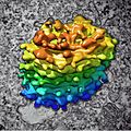 Algal Golgi body, 3D reconstruction (30466939665).jpg 2,043 × 2,043; 478 KB
-

-

-
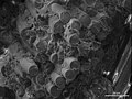
 Correlative 3D microscopy of plasmodesmata inside root nodules (25236962785).jpg 1,291 × 764; 193 KB
-
 Cross section of printed circuit board (25140841400).jpg 2,809 × 2,105; 436 KB
-
 Cultured Rat Hippocampal Neuron (24327909026).jpg 1,918 × 1,219; 784 KB
-
 Early human embryos (27872285595).jpg 1,300 × 1,030; 204 KB
-

-
-
-
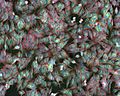
 Holmium oxide lamellar particles.png 1,024 × 768; 642 KB
-

-
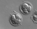
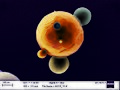
 Human embryos (four-cell stage) (27771482412).jpg 650 × 496; 58 KB
-
 Indian Muntjac fibroblast cells (23725924864).jpg 1,924 × 1,218; 718 KB
-
 Indian Muntjac fibroblast cells (24271618921).jpg 1,923 × 1,210; 684 KB
-
 Indian Muntjac fibroblast cells (24327908636).jpg 1,920 × 1,217; 1,007 KB
-
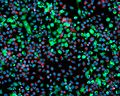
 Kidney section, fluorescence microscopy (30575642655).jpg 12,182 × 7,147; 6.17 MB
-
 Laser structured surface (24019830053).jpg 800 × 600; 335 KB
-
-
 Milled aluminum surface (24278900649).jpg 800 × 600; 260 KB
-
 Mitotic LLC-PK1 cells, fluorescence microscopy (23700644352).jpg 1,800 × 1,200; 1.6 MB
-
 Mobile phone camera lens module, 3D X-ray microscopy (30033111412).jpg 8,592 × 7,823; 7.56 MB
-
 Mouse Intestine (23727287703).jpg 1,915 × 1,217; 1,010 KB
-
 Mouse Kidney (23725924684).jpg 1,916 × 1,210; 993 KB
-
 Mouse tissue, stained histology preparation (23180949464).jpg 4,248 × 2,832; 2.19 MB
-
 Mouse tissue, stained histology preparation (23782972906).jpg 4,248 × 2,832; 1.97 MB
-
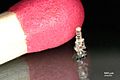 Nanosize 3D model of a vintage ZEISS microscope, by Nanoscribe (29829841445).jpg 1,457 × 970; 206 KB
-
 Nanoworlds (29746468381).jpg 2,048 × 1,536; 1.18 MB
-
 Oocyte with Zona pellucida (27771482282).jpg 1,300 × 1,030; 153 KB
-

-
 Rat primary cortical neuron culture, 3D reconstruction (30614936992).jpg 1,643 × 1,052; 629 KB
-
 Rat primary cortical neuron culture, deconvolved z-stack overlay (30614937102).jpg 2,752 × 2,208; 1.86 MB
-
-
-
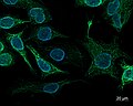 SK8-18-2 human derived cells, fluorescence microscopy (29942101073).jpg 2,758 × 2,214; 450 KB
-
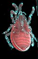
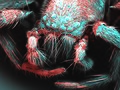 Spider, false-coloured scanning electron micrograph (27813345024).jpg 1,024 × 768; 1.06 MB
-
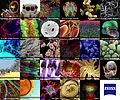 ZEISS and Cell Press present the Cell Picture Show (14989291035).jpg 1,800 × 1,500; 2.67 MB
.jpg)
.jpg)
.jpg)
.jpg)
.jpg)
.jpg)
.jpg)
.jpg)
.jpg)
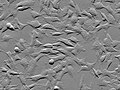
_cells,_phase_contrast_microscopy_with_ZEISS_Celldiscoverer_7_(30614936902).jpg)
.jpg)

_(27771482412).jpg)
.jpg)
.jpg)
.jpg)
.jpg)
.jpg)
.jpg)
.jpg)
.jpg)
.jpg)
.jpg)
.jpg)
.jpg)
.jpg)
.jpg)
.jpg)
.jpg)
.jpg)
.jpg)
.jpg)
.jpg)
.jpg)
.jpg)
.jpg)
.jpg)
.jpg)