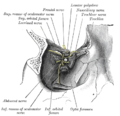
Category:Superior orbital fissure
Jump to navigation
Jump to search
foramen in the skull allowing for passage of cranial nerves, the ophthalmic vein, and sympathetic nerves | |||||
| Upload media | |||||
| Instance of |
| ||||
|---|---|---|---|---|---|
| Subclass of |
| ||||
| Part of |
| ||||
| |||||
Media in category "Superior orbital fissure"
The following 22 files are in this category, out of 22 total.
-
'Model, Sphenoid Bone, Free-standing' by William Rush.JPG 3,124 × 2,128; 532 KB
-
Braus 1921 338.png 1,612 × 908; 4.2 MB
-
Braus 1921 339.png 1,572 × 924; 4.16 MB
-
Cranial nerve foramina within middle cranial fossa.png 1,894 × 669; 1.95 MB
-
Gray145.png 724 × 446; 51 KB
-
Gray147.png 600 × 404; 39 KB
-
Gray787.png 413 × 400; 36 KB
-
Orbita mensch.jpg 500 × 374; 64 KB
-
Orbita1.jpg 960 × 720; 96 KB
-
Orbital cavity.jpg 960 × 720; 98 KB
-
Orbital fissures schematic.png 500 × 500; 32 KB
-
Skull foramina labeled ja.svg 1,089 × 957; 1.04 MB
-
Skull foramina labeled vie.svg 1,334 × 951; 1,024 KB
-
Skull foramina labeled.svg 1,334 × 951; 1,023 KB
-
Slide2hal.JPG 960 × 720; 93 KB
-
Slide3hal.JPG 960 × 720; 72 KB
-
Sobo 1909 53.png 2,304 × 1,288; 8.51 MB
-
Sobo 1909 54.png 2,232 × 1,344; 8.6 MB
-
Sobo 1909 95.png 1,716 × 1,252; 1.34 MB
-
Sobo 1909 98.png 2,308 × 1,476; 2.74 MB
-
Sphenoid bone - anterior view.jpg 960 × 720; 87 KB
-
Superior orbital fissure.PNG 411 × 626; 661 KB


