Category:Standard Event System
Jump to navigation
Jump to search
English: The "Standard Event System" (SES) to Study Vertebrate Embryos was developed in 2009 to establish a common language in comparative embryology
guideline to study vertebrate embryos | |||||
| Upload media | |||||
| |||||
Media in category "Standard Event System"
The following 110 files are in this category, out of 110 total.
-
 AA1. Trunk coiling (M02a).png 489 × 396; 103 KB
-
 AB1. Marsupium anlage (M03a).png 491 × 228; 139 KB
-
 AB2. Marsupium dent (M03b).png 487 × 214; 79 KB
-
 AC1. Monotreme beak (MO01a).png 478 × 232; 149 KB
-
 AD1. Hemipenes appears (Sq01a).png 187 × 142; 10 KB
-
 AD2. Hemipenes disappeared (Sq01b).png 191 × 142; 11 KB
-
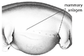 AE1. Mammary anlage (M04a).png 716 × 477; 234 KB
-
 AF1. Tongue protruding (T02a).png 464 × 301; 51 KB
-
 AG1. Plagiopatagium (M05a).png 826 × 839; 104 KB
-
 C1. Primitive streak present (V03a).png 481 × 218; 46 KB
-
 C2. Neural folds begin to close (V03b).png 519 × 194; 38 KB
-
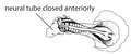 C3. Anterior neuropore closed (V03c).png 479 × 199; 38 KB
-
 C4. Posterior neuropore closed (V03d).png 463 × 192; 42 KB
-
 Character A1. Egg lay (V01a).png 481 × 235; 72 KB
-
 Character B1. Blastoporus (V02a).png 485 × 200; 40 KB
-
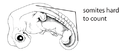 Character D1. Somites hard to count (V04a).png 491 × 215; 45 KB
-
 D2ff. somite pairs (V04b ff).png 486 × 214; 38 KB
-
 E1. Head bulbus (V05a).png 488 × 243; 62 KB
-
 E2. Anterior cephalic projection (V05b).png 484 × 207; 40 KB
-
 E3. Head projection disappeared (V05c).png 487 × 250; 52 KB
-
 F1. Olfactory placode (V06a).png 476 × 207; 33 KB
-
 F2. External nares (V06b).png 483 × 253; 30 KB
-
 F3. Nostrils formed (V06c).png 694 × 493; 153 KB
-
 Forelimb ridge (V12a).png 483 × 186; 11 KB
-
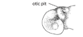 G1. Otic placode (V07a).png 480 × 214; 33 KB
-
 G2. Otic vesicle (V07b).png 489 × 239; 37 KB
-
 G3. Otic capsule inconspicuous (V07c).png 490 × 225; 51 KB
-
 G4. Pinna fold (V07d).png 249 × 166; 43 KB
-
 H1. Optic vesicle (V08a).png 461 × 232; 39 KB
-
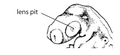 H2. Lens placode (V08b).png 476 × 217; 46 KB
-
 H4. Contour lens-iris (V08d).png 486 × 228; 44 KB
-
 H5. Pupil forms (V08e).png 484 × 239; 36 KB
-
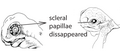 H7. Scleral papillae inconspicuous (V08g).png 480 × 217; 48 KB
-
 I1. Rib primordial visible (V09a).png 482 × 232; 48 KB
-
 J1. Ventricle bulbus (V10a).png 453 × 228; 43 KB
-
 J2. Thoracal bulbus disappeared (V10b).png 458 × 205; 58 KB
-
 J3. Ventricle S-shaped (V10c).png 429 × 188; 45 KB
-
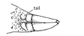
 K1. Tail bud (V11a).png 486 × 235; 54 KB
-
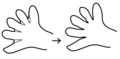
 L11. Forelimb paddle (V12g).png 484 × 102; 13 KB
-
 L13. Forelimb digital plate (V12i).png 466 × 112; 15 KB
-
 L15. Digital grooves forelimb (V12k).png 461 × 109; 12 KB
-
 L17. Digital serration forelimb (V12l).png 466 × 87; 11 KB
-
 L19. Finger (V12m).png 353 × 91; 8 KB
-
 L20. Toe (V12w).png 279 × 91; 6 KB
-
 L21. First claw forelimb (V12n).png 466 × 105; 12 KB
-
 L23. Hind limb sporn (V12q).png 481 × 121; 19 KB
-
 L25. Membranes between the fingers are completely gone (V12s).png 856 × 422; 12 KB
-
 L27. Separate phalanges are visible in the fingers (V12u).png 805 × 364; 156 KB
-
 L3. Forelimb bud (V12b).png 318 × 91; 9 KB
-
 L5. Forelimb elongated (V12c).tif 502 × 117; 17 KB
-
 L7. Forelimb AER (V12d).png 486 × 114; 14 KB
-
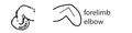 L9. Forelimb elbow (V12f).png 480 × 120; 16 KB
-
 M1. Head scales (V13a).png 476 × 207; 32 KB
-
 M10. Tail scales (V13h).png 462 × 199; 28 KB
-
 M11. Carapace scales (V13i).png 469 × 239; 44 KB
-
 M2. Throat scales (V13b).png 364 × 146; 27 KB
-
 M3. Lower eyelid scales (V13c).png 398 × 138; 22 KB
-
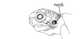 M4. Neck scales (V13d).png 473 × 221; 31 KB
-
 M5. Back scales (V13e).png 473 × 214; 49 KB
-
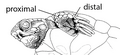 M6. Forelimb scales proximal (V13f).png 779 × 360; 120 KB
-
 M8. Hind limb scales proximal (V13f).png 618 × 627; 104 KB
-
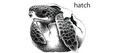 N1. Hatch (V14a).png 473 × 210; 82 KB
-
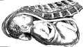 N2. Birth (V14b).png 297 × 170; 4 KB
-
 N3. Leaving pouch (V14c).jpg 189 × 218; 13 KB
-
 N4. Weaning (V14d).gif 297 × 222; 3 KB
-
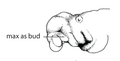 O1. Maxillary bud (G01a).png 466 × 237; 36 KB
-
 O2. Maxillary process posterior to eye (G01b).png 476 × 217; 52 KB
-
 O3. Maxillary process midline of eye (G01c).png 459 × 219; 35 KB
-
 O4. Maxillary process anterior to lens (G01d).png 461 × 192; 52 KB
-
 O5. Maxillary process anterior to eye (G01e).png 480 × 214; 44 KB
-
 O6. Maxillary process posterior to eye (G01f).png 458 × 232; 34 KB
-
 P1. Mandibular arch bud (G02a).png 476 × 192; 40 KB
-

-
 P3. Mandibular arch bud posterior lens (G02c).png 476 × 185; 30 KB
-
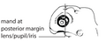
 P4. Mandibular arch bud midline eye (G02d).png 476 × 199; 33 KB
-
 P5. Mandibular arch bud anterior lens (G02e).png 469 × 210; 42 KB
-
 P6. Mandibular arch bud anterior eye (G02f).png 476 × 204; 38 KB
-
 P7. Mandibular arch bud level frontonasal groove (G02g).png 577 × 252; 51 KB
-
 P8. Mandibular arch bud occlusion point = jaw closure (G02h).png 469 × 207; 41 KB
-
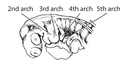 Q1. Second arch bud (G03a).png 478 × 261; 68 KB
-
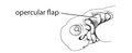 Q2. Hyoid flap (G03e).png 480 × 196; 27 KB
-
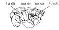 R1. First pharyngeal slit (G04a).png 461 × 239; 59 KB
-
 R5. Pharyngeal slits closed (G04e).png 473 × 199; 37 KB
-
 S1. Urogenital papilla bud (G05a).png 480 × 180; 28 KB
-
 S2. Urogenital papilla bud inconspicuous (G05b).png 466 × 207; 44 KB
-
 Standard Event System.png 1,694 × 1,682; 1.5 MB
-
 T1. Cervical flexure 90° (T01a).png 423 × 219; 35 KB
-
 T2. Cervical flexure disappeared (T01b).png 486 × 221; 63 KB
-
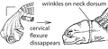
 T4. Nuchal fold (T01d).png 519 × 267; 42 KB
-
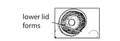 U1. Lower lid appears (A01a).png 445 × 152; 26 KB
-
 U2. Eyelid begun overgrow (A01b).png 454 × 145; 29 KB
-
 U3. Eyelid at level of scleral papillae (A01c).png 441 × 145; 36 KB
-
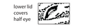
 U4. Eyelid ventral to lens (A01d).png 454 × 149; 40 KB
-
 U5. Eyelid covers eye (A01e).png 452 × 149; 26 KB
-
 U6. Membrana nictitans (A01f).png 445 × 160; 26 KB
-
 U7. Eyelid open again (A01g).png 1,278 × 905; 230 KB
-
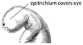 U8. Epitrichium covering eye (A01h).png 1,098 × 595; 223 KB
-
 V1. Caruncle appears (A02a).png 809 × 517; 116 KB
-
 W1. Ramphothecae appear (S01a).png 448 × 225; 30 KB
-
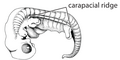 X1. Carapacial ridge (S02a).png 445 × 221; 59 KB
-
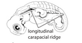 X2. Longitudinal carapacial ridge (S02b).png 454 × 239; 46 KB
-
 X3. Carapace not reaching anterior (S02c).png 458 × 225; 54 KB
-
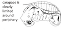 X4. Carapace clearly limited (S02d).png 458 × 228; 52 KB
-
 X5. Carapace posterior projection (S02e).png 462 × 246; 58 KB
-
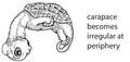 X6. Carapace irregular (S02f).png 449 × 212; 51 KB
-
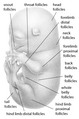 Y1. Hair follicles (M04).png 543 × 818; 316 KB
-
 Z1. Back hairs (M01a).png 484 × 262; 59 KB
-
 Z4. Forelimb hairs proximal (M01d).png 475 × 243; 67 KB
-
 Z6. Hind limb hairs proximal (M01g).png 484 × 233; 43 KB
-
 Z9. Neck hairs (M01i).png 404 × 465; 128 KB
.png)
.png)
.png)
.png)
.png)
.png)
.png)
.png)
.png)
.png)
.png)
.png)
.png)
.png)
.png)
.png)
.png)
.png)
.png)
.png)
.png)
.png)
.png)
.png)
.png)
.png)
.png)
.png)
.png)
.png)
.png)
.png)
.png)
.png)
.png)
.png)
.png)
.png)
.png)
.png)
.png)
.png)
.png)
.png)
.png)
.png)
.jpg)
.gif)
.png)
.png)
.png)
.png)
.png)
.png)
.png)
.png)
.png)
.png)
.png)
.png)
.png)
.png)
.png)
.png)

.png)
.png)
.png)
.png)
.png)
.png)
.png)
.png)
.png)
.png)
.png)
.png)
.png)
.png)
.png)
.png)
.png)
.png)
.png)
.png)
.png)
.png)
.png)
.png)
.png)
.png)
.png)
.png)
.png)
.png)
.png)
.png)
.png)
.png)
.png)
.png)
.png)
.png)
.png)
.png)
.png)
.png)
.png)
.png)