Category:Sphenoid wing meningioma Case 001
Jump to navigation
Jump to search
| This image set can be scrolled interactively with the mouse (wheel or dragging). This is done by using the template Imagestack. |
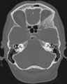
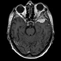
Deutsch: Keilbeinmeningeom in der Computertomographie und Magnetresonanztomographie. Deutliche, homogene Kontrastmittelaufnahme mit 'dural tail' (sagittal gut zu sehen). In der Computertomographie (Knochenfenster und VRT) ist die reaktive Hyperostose besonders gut zu erkennen, die durch die Vorwölbung in die Orbita schon einen Bulbustiefstand bewirkte.


Impression of the left orbita by hyperostosis
Media in category "Sphenoid wing meningioma Case 001"
The following 18 files are in this category, out of 18 total.
-
 Keilbeinmeningeom CT ax.png 812 × 1,018; 357 KB
-
 Keilbeinmeningeom CT cor.png 920 × 942; 287 KB
-
 Keilbeinmeningeom CT VRT.png 800 × 868; 720 KB
-
 Keilbeinmeningeom CT-VRT-0.jpg 512 × 512; 65 KB
-
 Keilbeinmeningeom CT-VRT-1.jpg 512 × 512; 63 KB
-
 Keilbeinmeningeom CT-VRT-2.jpg 512 × 512; 61 KB
-
 Keilbeinmeningeom CT-VRT-3.jpg 512 × 512; 59 KB
-
 Keilbeinmeningeom CT-VRT-4.jpg 512 × 512; 57 KB
-
 Keilbeinmeningeom CT-VRT-5.jpg 512 × 512; 58 KB
-
 Keilbeinmeningeom CT-VRT-6.jpg 512 × 512; 60 KB
-
 Keilbeinmeningeom CT-VRT-7.jpg 512 × 512; 62 KB
-
 Keilbeinmeningeom CT-VRT-8.jpg 512 × 512; 64 KB
-
 Keilbeinmeningeom CT-VRT-9.jpg 512 × 512; 66 KB
-
 Keilbeinmeningeom MRT T1ax.jpg 958 × 961; 78 KB
-
 Keilbeinmeningeom MRT T1KMax.jpg 958 × 961; 79 KB
-
 Keilbeinmeningeom MRT T1KMcor.png 774 × 984; 296 KB
-
 Keilbeinmeningeom MRT T1KMsag.png 938 × 988; 285 KB
-
 Keilbeinmeningeom MRT T2ax.jpg 958 × 961; 106 KB


