Category:Rodentia anatomy
Jump to navigation
Jump to search
English: +/−
Subcategories
This category has the following 25 subcategories, out of 25 total.
C
D
E
- Erethizontidae anatomy (6 F)
G
H
- Heteromyidae anatomy (5 F)
M
O
- Octodon degus anatomy (18 F)
P
S
Media in category "Rodentia anatomy"
The following 22 files are in this category, out of 22 total.
-
3D image of mouse intestine (50069993586).jpg 706 × 496; 50 KB
-
AR-PAM Mouse Ear Vasculature.jpg 495 × 390; 32 KB
-
Callystomis pictus 1 - CMARF-UESC 1230.png 1,040 × 780; 550 KB
-
Callystomis pictus 2 - CMARF-UESC 1230.png 1,040 × 780; 460 KB
-
Demonstration of Cheek Pouches in Geomyid.jpg 303 × 540; 97 KB
-
Development of the respiratory system in mice..jpg 600 × 343; 135 KB
-
Etiqueta do exemplar de Callystomis pictus - CMARF-UESC 1230.png 1,040 × 780; 494 KB
-
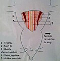
Localisation des vaisseaux.JPG 1,641 × 1,656; 699 KB
-
Lower Jaw in Rodents.png 2,432 × 1,534; 273 KB
-
Maus-skelett hg.jpg 3,433 × 2,562; 1.12 MB
-
Mouse anatomy for scientific publication.svg 159 × 177; 107 KB
-
Mouse ISH coronal slide, Slc6a3, substantia nigra.jpg 10,657 × 7,153; 20.48 MB
-
Mouse whole-mount meninges.png 552 × 643; 1.39 MB
-
Nagethiere plate LIV.jpg 2,140 × 2,572; 1.59 MB
-
Nagethiere plate LV.jpg 2,136 × 2,568; 1.3 MB
-
Nagethiere plate LVI.jpg 2,106 × 2,584; 1.15 MB
-
Nagethiere plate LVII.jpg 2,112 × 2,580; 1.3 MB
-
OR-PAM Mouse Ear Vasculature.jpg 1,050 × 527; 78 KB
-
Rodent Teeth, Hunterian Museum.png 366 × 513; 128 KB
-
The primary dental lamina.jpg 600 × 210; 44 KB


