Category:Porifera diagrams
Jump to navigation
Jump to search
Media in category "Porifera diagrams"
The following 55 files are in this category, out of 55 total.
-
 Ascon anatomia.jpg 1,499 × 1,761; 237 KB
-
 Ascon body wall.PNG 417 × 541; 19 KB
-
 Ascon.jpg 151 × 304; 32 KB
-
 Ascon.PNG 221 × 308; 12 KB
-
 Asconoid Sponge body plan.png 1,068 × 1,232; 300 KB
-
 Asconoide-fr.svg 1,500 × 1,800; 150 KB
-
 Asconoide-it.svg 1,500 × 1,800; 148 KB
-
 Askon.jpg 208 × 295; 16 KB
-
 Bathymetrical range of selected sponge species.jpg 801 × 1,200; 75 KB
-
 Choanoflagellate and choanocyte.png 1,030 × 515; 233 KB
-
 Diagram Bekerspons.png 1,350 × 1,558; 785 KB
-
 EB1911 Sponges - Plakina monclopha (1).jpg 896 × 1,076; 530 KB
-
 Esponjas alimentacion.jpg 2,447 × 1,287; 268 KB
-
 Euplectella 01.png 180 × 247; 4 KB
-
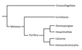
 Filogenia porifera.jpg 852 × 995; 190 KB
-
 Global diversity of the Porifera - ru.png 2,048 × 1,117; 851 KB
-
 Global diversity of the Porifera.png 2,048 × 1,117; 856 KB
-
 Ishrana.PNG 783 × 412; 47 KB
-
 Leukon.jpg 183 × 251; 10 KB
-
 Meyers b14 s0680a.jpg 1,448 × 2,343; 2.9 MB
-
 Perrier 948389.jpg 2,544 × 3,304; 2.28 MB
-
 Phylogénie Porifera.png 1,156 × 702; 17 KB
-
 Porifera body structures 01.png 1,316 × 700; 287 KB
-
 Porifera calcifying 01.png 226 × 206; 5 KB
-
 Porifera cell types 01.png 197 × 232; 4 KB
-
 Porifera diversity.png 997 × 917; 164 KB
-
 Porifera Types-fr.svg 1,310 × 770; 309 KB
-
 Porifera, Otto's Encyclopedia.jpg 1,478 × 1,876; 667 KB
-
 Porifera- Generalized Amphiblastula Larva Settling.jpg 6,523 × 6,937; 4.23 MB
-
 Poriferan phylogeny.PNG 497 × 307; 8 KB
-
 Prianosin D 2D.JPG 662 × 559; 30 KB
-
 PSM V03 D557 United spicules in pheronema.jpg 1,497 × 683; 41 KB
-
 PSM V03 D559 Extremity of a mooring thread of pheronema.jpg 1,375 × 277; 25 KB
-
 PSM V03 D559 Outer wall of ventriculites simplex.jpg 891 × 876; 144 KB
-
 PSM V03 D561 Outer surface of ventriculites simplex.jpg 845 × 768; 205 KB
-
 Schematic drawing of Amphiblastula type of development Calcarea.svg 1,048 × 592; 439 KB
-
 Schematic drawing of Calciblastula type of development Calcarea Calcinea.svg 1,056 × 697; 373 KB
-
 Schematic drawing of direct development Demospongiae Spirophorida.svg 1,034 × 717; 362 KB
-
 Schematic drawing of Disphaerula type of development Demospongiae.svg 1,000 × 676; 278 KB
-
 Schematic drawing of the first subtype of Parenchymella type of development.svg 1,058 × 818; 428 KB
-
 Schematic drawing of Trichimella type of development Hexactinellida - ru.svg 1,000 × 941; 448 KB
-
 Sea sponge diagram.svg 1,080 × 1,260; 709 KB
-
 Sea sponge.svg 1,080 × 1,260; 733 KB
-
 Sikon anatomy.jpg 1,612 × 842; 203 KB
-
 Sikon.PNG 751 × 344; 30 KB
-
 Spicule life cycle.jpg 1,030 × 1,200; 235 KB
-
 Spicule synthesis.PNG 1,061 × 238; 18 KB
-
 Spikule.PNG 525 × 852; 41 KB
-
 Sponge Cell Layers xs.jpg 756 × 265; 97 KB
-
 Sponge cells and spicule.png 1,500 × 843; 121 KB
-
 Sponge Cells.jpg 683 × 375; 55 KB
-
 Sponge spicule diversity.png 1,500 × 481; 98 KB
-
 Sponge whole.jpg 443 × 322; 40 KB
-
 Stylocordyla longissima.jpg 392 × 699; 40 KB
-
 Sykon.jpg 213 × 279; 11 KB


.jpg)


