Category:Plant cell diagrams
Jump to navigation
Jump to search
Subcategories
This category has the following 2 subcategories, out of 2 total.
S
- Stoma diagrams (22 F)
Pages in category "Plant cell diagrams"
This category contains only the following page.
Media in category "Plant cell diagrams"
The following 125 files are in this category, out of 125 total.
-
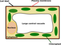
 2Plant cell structure.png 753 × 599; 201 KB
-
 A la estela.svg 3,000 × 1,420; 1.13 MB
-
 Antheridia Dutch text.jpg 400 × 300; 25 KB
-
 Apoplast and symplast pathways (id).jpg 800 × 566; 191 KB
-
 Apoplast and symplast pathways.png 466 × 249; 8 KB
-
 Apoplast and symplast pathways.svg 1,052 × 744; 16 KB
-
 Arabidopsis meristem WUS.png 700 × 415; 50 KB
-
 Archegonium Dutch text.jpg 294 × 300; 17 KB
-
 Biljke.jpg 410 × 444; 88 KB
-
 Botany (Page 34) BHL16936935.jpg 2,184 × 3,574; 1.31 MB
-
 Cadres de Caspary.JPG 859 × 605; 94 KB
-
 Celula vegetal.png 550 × 439; 597 KB
-
 Chloroplast diagram (zh-cn).svg 920 × 680; 27 KB
-
 Chloroplast diagram sr.svg 1,052 × 744; 23 KB
-
 Chloroplast en.png 744 × 438; 21 KB
-
 Chloroplast movement fr.svg 455 × 314; 79 KB
-
 Chloroplast movement id.svg 460 × 330; 195 KB
-
 Chloroplast movement pt.svg 460 × 330; 90 KB
-
 Chloroplast movement ua.svg 512 × 367; 43 KB
-
 Chloroplast movement.svg 460 × 330; 178 KB
-
 Chloroplast pl.png 748 × 501; 314 KB
-
 Chloroplast-new-Dutch.jpg 748 × 501; 66 KB
-
 Chloroplast-new.jpg 748 × 501; 54 KB
-
 Cyanogene Glukoside-fr.svg 600 × 350; 42 KB
-
 Cyanogene Glukoside.svg 600 × 350; 41 KB
-
 Development of Chloroplast.png 744 × 438; 8 KB
-
 Die Gartenlaube (1853) b 259.jpg 602 × 943; 161 KB
-
 Discontinuous Casparian strip in sgn3 mutant.jpg 852 × 607; 62 KB
-
 Disegno della cellula vegetale.jpg 2,044 × 3,251; 988 KB
-
 Drewno Dutch txt.jpg 400 × 384; 34 KB
-
 Drewno.jpg 400 × 384; 18 KB
-
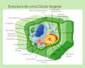
 Estructura celula vegetal.png 1,024 × 815; 297 KB
-
 Estructura cèl·lula vegetal.png 1,024 × 815; 289 KB
-
 Eukaryota cell strucutre.PNG 580 × 440; 148 KB
-
 Glyoxysome.jpg 454 × 424; 49 KB
-
 Golgi body.JPG 487 × 181; 22 KB
-
 Guard-cell-plant.png 1,297 × 769; 504 KB
-
 Kasvisolu.png 800 × 800; 300 KB
-
 Komorkaroslinna.png 902 × 774; 219 KB
-
 Komorkaroslinna.svg 512 × 450; 66 KB
-
 Magnesium in plant cell.png 487 × 503; 26 KB
-
 Magnesium in plant cell.svg 987 × 1,020; 41 KB
-
 Meyers b1 s0315 b1.png 240 × 120; 9 KB
-
 Meyers b1 s0315 b2.png 240 × 120; 7 KB
-
 Miękisz.jpg 400 × 375; 28 KB
-
 Morfoanatomia celula vegetal.png 649 × 475; 128 KB
-
 Opening and Closing of Stoma-bn.svg 1,297 × 769; 195 KB
-
 Parede celular vegetal.png 772 × 666; 69 KB
-
 Phloem Cells.jpg 274 × 522; 36 KB
-
 Phloem cells.svg 440 × 480; 4.38 MB
-
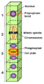
 Phragmoplast ru.png 585 × 396; 30 KB
-
 Phragmoplast.png 585 × 396; 11 KB
-
 Phragmosome svg ru.png 369 × 467; 49 KB
-
 Phragmosome.svg 369 × 467; 33 KB
-
 Phycoplast ru.png 443 × 434; 39 KB
-
 Phycoplast.png 443 × 434; 15 KB
-
 Plant cell early anaphase.svg 580 × 380; 91 KB
-
 Plant cell first gap.svg 580 × 400; 20 KB
-
 Plant cell late anaphase.svg 580 × 380; 105 KB
-
 Plant cell metaphase.svg 580 × 380; 86 KB
-
 Plant cell prometaphase.svg 580 × 380; 68 KB
-
 Plant cell prophase.svg 580 × 380; 74 KB
-
 Plant cell S phase and second gap.svg 580 × 385; 41 KB
-
 Plant cell showing primary and secondary wall by CarolineDahl.jpg 3,015 × 2,236; 878 KB
-
 Plant cell structure bn.png 753 × 599; 206 KB
-
 Plant cell structure Icelandic text.png 854 × 615; 276 KB
-
 Plant cell structure it.png 1,024 × 815; 291 KB
-
 Plant cell structure ku.svg 660 × 478; 186 KB
-
 Plant cell structure no text-2 small.svg 459 × 344; 99 KB
-
 Plant cell structure no text.png 1,024 × 815; 243 KB
-
 Plant cell structure pt.gif 1,024 × 815; 118 KB
-
 Plant cell structure pt.svg 649 × 475; 136 KB
-
 Plant cell structure sk.png 1,024 × 815; 66 KB
-
 Plant cell structure sr.PNG 753 × 599; 185 KB
-
 Plant cell structure-fr.png 1,024 × 815; 330 KB
-
 Plant cell structure-kn.png 649 × 475; 143 KB
-
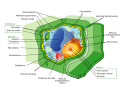
 Plant cell structure-ru-v1.png 900 × 740; 288 KB
-
 Plant cell structure-ru-v2.png 2,601 × 1,764; 920 KB
-
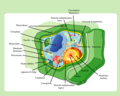
 Plant cell structure.png 1,024 × 815; 293 KB
-
 Plant Cell.jpg 1,031 × 612; 274 KB
-
 Plant Cell.png 649 × 475; 148 KB
-
 Plant sperm nb.png 367 × 656; 43 KB
-
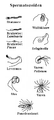 Plant sperm nl txt.png 383 × 852; 42 KB
-
 Plant sperm.png 301 × 602; 31 KB
-
 Plantcel.gif 175 × 287; 4 KB
-
 Plantcell2.png 650 × 478; 55 KB
-
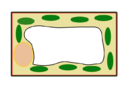 Plantcell2notext.png 650 × 478; 37 KB
-
 Plantencel structuur.PNG 753 × 599; 185 KB
-
 Plantencel.png 1,854 × 1,590; 1.14 MB
-
 Planzenzelle beschriftet.svg 512 × 404; 317 KB
-
 Plasmodesma.Int.png 459 × 367; 10 KB
-
 Preprophaseband ru.png 314 × 599; 73 KB
-
 Preprophaseband.png 412 × 786; 23 KB
-
 Pressurebomb (es).svg 394 × 274; 30 KB
-
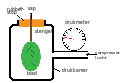 Pressurebomb Dutch txt.svg 2,000 × 1,391; 153 KB
-
 Pressurebomb ml.svg 394 × 274; 108 KB
-
 Pressurebomb-hi.svg 394 × 274; 107 KB
-
 Pressurebomb-kn.svg 394 × 274; 100 KB
-
 Pressurebomb-mr.svg 394 × 274; 107 KB
-
 Pressurebomb-pa.svg 394 × 274; 109 KB
-
 Pressurebomb-te.svg 394 × 274; 104 KB
-
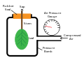 Pressurebomb.svg 394 × 274; 100 KB
-
 PSM V21 D307 Cross section of petiole with crystals in the cells.jpg 1,220 × 1,131; 380 KB
-
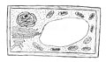
 PSM V30 D195 Testing energy of plant cells.jpg 689 × 1,242; 96 KB
-
 Scholander-Bombe austretender Tropfen.JPG 3,008 × 2,000; 1.45 MB
-
 Scholander-Bombe DSC 8061.JPG 3,008 × 2,000; 2.56 MB
-
 Schéma pression osmotique cellule végétale.png 927 × 366; 79 KB
-
 SieveTube.png 120 × 317; 7 KB
-
 Simple Pit Pair.jpg 800 × 600; 19 KB
-
 Symplastic and apoplastic water flow through root (TC ver).png 1,462 × 910; 5.1 MB
-
 Symplastic and apoplastic water flow through root.png 1,502 × 1,062; 142 KB
-
 Turgescence-et-plasmolyse-cellule-vegetale.png 952 × 568; 70 KB
-
 Turgor pressure on plant cells diagram pl.svg 619 × 272; 99 KB
-
 Turgor pressure on plant cells diagram-it.svg 618 × 244; 53 KB
-
 Twardzica.jpg 80 × 154; 6 KB
-
 Untitled drawing (1).pdf 1,500 × 1,125; 43 KB
-
 Vakuol i Växtcell.png 507 × 242; 20 KB
-
 Xaneya riwekan .png 693 × 508; 163 KB
-
 Xaneya riwekan ku.svg 692 × 507; 128 KB
-
 Xylem Cell.jpg 231 × 467; 47 KB
-
 Xylem cells ta.png 600 × 705; 221 KB
-
 Zwarcica.jpg 450 × 171; 29 KB
-
 植物細胞.png 919 × 660; 64 KB
-
 植物細胞ゴルジ体.png 919 × 660; 37 KB
-
 植物細胞葉緑体.png 919 × 660; 42 KB


.jpg)


_BHL16936935.jpg)


.svg)


_b_259.jpg)


.svg)


.png)


