Category:Optic chiasm
Jump to navigation
Jump to search
optical part of brain | |||||
| Upload media | |||||
| Instance of |
| ||||
|---|---|---|---|---|---|
| Subclass of |
| ||||
| Part of | |||||
| |||||
Media in category "Optic chiasm"
The following 68 files are in this category, out of 68 total.
-
1204 Optic Nerve vs Optic Tract.jpg 787 × 615; 215 KB
-
1543,Vesalius'Fabrica,VisualSystem,V1.jpg 286 × 309; 25 KB
-
1543,Visalius'OpticChiasma.jpg 287 × 308; 24 KB
-
Acromegaly pituitary macroadenoma.JPEG 905 × 452; 89 KB
-
Anatomy of the cavernous sinus.jpg 800 × 499; 93 KB
-
AxialTwistDevelopment.png 763 × 1,220; 479 KB
-
AxialTwistScenario.png 642 × 310; 132 KB
-
B112px-1543,Visalius'OpticChiasma.jpg 50 × 50; 1 KB
-
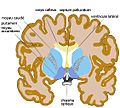
Basal ganglia.jpg 564 × 507; 54 KB
-
Brain MRI 150443 rgbca t1 t2 t2STIR misreg.png 419 × 522; 493 KB
-
Caudal view of a brain after dissection.png 1,890 × 2,405; 2.94 MB
-
Cavernous malformation of the optic chiasm.jpg 600 × 450; 297 KB
-
Constudeyepath el.png 448 × 589; 72 KB
-
Constudeyepath ja.png 800 × 1,139; 112 KB
-
Constudeyepath.png 501 × 650; 13 KB
-
Contralateral brain.pdf 337 × 789; 6 KB
-
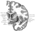
Gray1180-ar.png 672 × 375; 165 KB
-
Gray1180.png 672 × 375; 45 KB
-
Gray722 ar.png 360 × 500; 705 KB
-
Gray722-ar.svg 463 × 543; 504 KB
-
Gray722-svg (zh-tw).svg 463 × 543; 95 KB
-
Gray722-svg-de.svg 463 × 543; 116 KB
-
Gray722-svg-FR.svg 463 × 543; 95 KB
-
Gray722.png 360 × 500; 19 KB
-
Gray722.svg 463 × 543; 103 KB
-
Gray722ita.png 360 × 500; 19 KB
-
Gray744.png 550 × 481; 54 KB
-
Gray748.png 500 × 510; 66 KB
-
Gray773.png 392 × 450; 22 KB
-
Gray774 he.png 402 × 550; 175 KB
-
Gray774.png 402 × 550; 20 KB
-
Grays pituitary.png 600 × 333; 65 KB
-
Hemia ita.PNG 624 × 624; 65 KB
-
Hemia.PNG 624 × 624; 64 KB
-
Hemianopsien.png 624 × 624; 77 KB
-
Human brain anterior-inferior view description.JPG 330 × 475; 31 KB
-
Human brainstem anterior view description 2.JPG 347 × 485; 31 KB
-
LocationOfHypothalamus.jpg 350 × 250; 21 KB
-
Mouse and human visual systems.jpg 850 × 731; 148 KB
-
Optic chiasm, human, 19th century. Wellcome L0002031.jpg 1,551 × 1,195; 732 KB
-
Optic nerve and ocular muscles. Wellcome L0009855.jpg 1,604 × 1,222; 861 KB
-
Optic nerve optic tract optic radiation.jpg 826 × 2,208; 933 KB
-
Optic nerve optic tract optic radiation2.jpg 1,418 × 1,923; 1.34 MB
-
Optic nerve pair & two brain hemispheres.jpg 252 × 405; 21 KB
-
Optic pathway.png 1,890 × 2,528; 1.29 MB
-
Optic processing human brain.jpg 501 × 700; 137 KB
-
Optic tract and optic nerve.jpg 960 × 720; 90 KB
-
Optic-cabling he.JPG 800 × 600; 53 KB
-
Optical-transformations.png 1,415 × 2,085; 411 KB
-
Pituitary gland-optic chiasm-sella turcica-es.png 625 × 347; 69 KB
-
Pituitary gland-optic chiasm-sella turcica.jpg 600 × 333; 127 KB
-
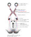
Schematic drawing of the human visual system.jpg 981 × 1,368; 381 KB
-
Sehbahn 3D v2.jpg 817 × 674; 94 KB
-
Sehbahn 3D.jpg 817 × 674; 99 KB
-
Sehbahn mit Chiasma opticum.jpg 864 × 1,080; 182 KB
-
Slide13qq.JPG 960 × 720; 89 KB
-
Slide2cuc.JPG 960 × 720; 101 KB
-
Slide2Dsa.JPG 960 × 720; 89 KB
-
Slide2HOM.JPG 960 × 720; 82 KB
-
Slide2ZEB.JPG 960 × 720; 106 KB
-
Slide3MIR.JPG 960 × 720; 127 KB
-
Slide4dd.JPG 960 × 720; 109 KB
-
Slide4MIR.JPG 960 × 720; 97 KB
-
Sobo 1909 649.png 816 × 520; 1.22 MB
-
Sobo 1911 747.png 1,964 × 1,080; 6.08 MB
-
Sysème visuel humain.jpg 787 × 615; 135 KB
-
Visualfield.jpg 353 × 377; 24 KB
-
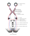
Voies visuelles3.svg 744 × 964; 151 KB


