Category:Mikael Häggström/X-rays of the upper extremity
Jump to navigation
Jump to search

These images were created by Mikael Häggström, M.D.
- User info
- Reusing images
Subcategories
This category has only the following subcategory.
Media in category "Mikael Häggström/X-rays of the upper extremity"
The following 22 files are in this category, out of 22 total.
-
 Calcinosis of CREST syndrome.jpg 554 × 605; 84 KB
-
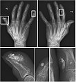
 Erosive osteoarthritis with gull-wing appearance.jpg 1,087 × 886; 170 KB
-
 Radiograph of Barton's fracture.jpg 319 × 612; 57 KB
-

-

-

-
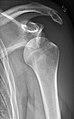 Shoulder dislocation, anteroposterior before reduction.jpg 647 × 1,041; 128 KB
-
 Shoulder dislocation, Y-projection after reduction.jpg 559 × 1,049; 134 KB
-
 Shoulder dislocation, Y-projection before reduction.jpg 561 × 965; 124 KB
-
 X-ray of dynamic scapholunate instability.jpg 847 × 690; 190 KB
-
 X-ray of normal elbow by 30 degrees internal oblique projection.jpg 693 × 1,135; 85 KB
-
 X-ray of normal elbow by anteroposterior projection.jpg 849 × 1,138; 98 KB
-
 X-ray of normal elbow by lateral projection.jpg 1,086 × 1,103; 115 KB
-
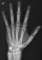 X-ray of normal hand by dorsoplantar projection.jpg 1,466 × 2,082; 493 KB
-
 X-ray of normal hand by lateral projection.jpg 686 × 2,123; 246 KB
-
 X-ray of normal hand by oblique projection.jpg 1,003 × 2,098; 379 KB
-
 X-ray of normal wrist by dorsoplantar projection (crop).jpg 690 × 980; 204 KB
-
 X-ray of normal wrist by dorsoplantar projection.jpg 690 × 2,122; 364 KB
-
 X-ray of normal wrist by lateral projection (crop).jpg 559 × 825; 95 KB
-
 X-ray of osteogenesis imperfecta type 5 in newborn - left arm - annotated.jpg 832 × 1,440; 148 KB
-
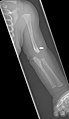
 X-ray of osteogenesis imperfecta type 5 in newborn - left arm.jpg 832 × 1,440; 147 KB
-
 X-ray of osteogenesis imperfecta type 5 in newborn - right arm.jpg 1,269 × 935; 108 KB


.jpg)
.jpg)


