Category:Mikael Häggström/Computed tomography images of the abdomen and pelvis
Jump to navigation
Jump to search

These images were created by Mikael Häggström, M.D.
- User info
- Reusing images
Subcategories
This category has the following 3 subcategories, out of 3 total.
M
Media in category "Mikael Häggström/Computed tomography images of the abdomen and pelvis"
The following 32 files are in this category, out of 32 total.
-
 Abdominal CT with scan range and field of view, with box and text.jpg 1,915 × 1,461; 607 KB
-
 Abdominal CT with scan range and field of view, with box and text.svg 979 × 747; 308 KB
-
 Abdominal CT with scan range and field of view, with box.jpg 979 × 747; 221 KB
-
 Abdominal CT with scan range and field of view, without annotations.jpg 929 × 727; 204 KB
-
 CT of a normal abdomen and pelvis, thumbnail.png 644 × 899; 445 KB
-
 CT of cholecystitis.jpg 914 × 720; 121 KB
-
 CT of peripelvic cysts with non-contrast and urography.jpg 650 × 404; 69 KB
-
 CT of rectus sheath hematomas, follow-up.png 675 × 860; 453 KB
-
 CT of rectus sheath hematomas.png 650 × 757; 374 KB
-
 CT urography of peripelvic cysts.jpg 1,005 × 1,009; 280 KB
-
 Interspinous distance, thin slice, annotated.jpg 977 × 498; 126 KB
-
 Low-dose CT of interspinous diameter.jpg 1,105 × 765; 341 KB
-
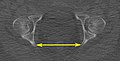
 Low-dose CT of intertuberous diameter.jpg 482 × 504; 86 KB
-
 Low-dose CT of obstetric conjugate and pelvic outlet.jpg 711 × 761; 162 KB
-
 Low-dose CT of obstetric conjugate.jpg 711 × 761; 156 KB
-
 Low-dose CT of sagittal pelvic outlet diameter.jpg 758 × 812; 168 KB
-

-
 Low-dose CT of transverse diameter of pelvic inlet.jpg 1,105 × 765; 343 KB
-
 Low-dose CT scan of interspinous distance, axial plane.jpg 995 × 775; 200 KB
-
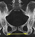
 Low-dose CT scan of intertuberous diameter, axial plane.jpg 430 × 304; 69 KB
-
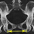 Low-dose CT scan of intertuberous diameter.jpg 504 × 504; 86 KB
-

-

-
 Non-contrast CT of peripelvic cysts.jpg 945 × 985; 185 KB
-
-
 Volume rendered CT scan of a pregnancy of 37 weeks of gestational age.gif 874 × 996; 49.91 MB
-
 Volume rendered CT scan of abdominal and pelvic blood vessels (smaller).gif 644 × 862; 45.23 MB
-
 Volume rendered CT scan of abdominal and pelvic blood vessels.gif 822 × 1,100; 62.98 MB
-
 Volume rendering of abdominal muscles.jpg 771 × 943; 219 KB
-
 Volume rendering of hepatic veins, annotated.jpg 1,784 × 1,606; 666 KB
-
 Volume rendering of hepatic veins, annotated.svg 892 × 803; 1.41 MB
-
 Volume rendering of hepatic veins.jpg 892 × 803; 241 KB


.gif)

.gif)


