
Category:Microscopic images relating to animals

Subcategories
This category has the following 19 subcategories, out of 19 total.
*
A
B
C
F
M
N
R
Media in category "Microscopic images relating to animals"
The following 134 files are in this category, out of 134 total.
-
 Ammonite fossils.JPG 5,184 × 3,456; 12.73 MB
-
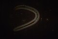
 An alien among the stars.jpg 1,080 × 1,620; 232 KB
-
 An omnivorous nematode in the laboratory.jpg 6,016 × 4,000; 9.33 MB
-
LL-Q1860 (eng)-Vealhurl-animalcule.wav 1.5 s; 139 KB
-
 Animalcules in the teeth Wellcome L0002254.jpg 1,152 × 1,598; 1,020 KB
-
 Animalcules in the teeth. Wellcome L0002253.jpg 1,190 × 1,546; 811 KB
-

-
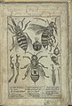 Apiario 5r top.jpg 889 × 1,313; 169 KB
-
 Ascariscs40x2.jpg 1,024 × 768; 184 KB
-
 Asterolepis tooth cross-section.jpg 280 × 261; 53 KB
-
 Biological cell.svg 1,466 × 891; 249 KB
-
 Bipinnaria Larvae of a developing starfish.jpg 3,024 × 4,032; 1.73 MB
-
 Bipinnaria of starfish.JPG 800 × 1,020; 257 KB
-
 Bipinnaria.jpg 1,620 × 2,392; 516 KB
-
 BloodFlukeEggs40x2.jpg 1,024 × 768; 130 KB
-
 Branchiostoma lanceolatum (cropped).jpg 701 × 679; 20 KB
-
 Branchiostoma lanceolatum.jpg 1,024 × 811; 28 KB
-
 Brehms Tierleben. Allgemeine kunde des Tierreichs (1918) (20227843139).jpg 2,198 × 3,098; 1.78 MB
-
 C wegeneri.JPG 648 × 486; 284 KB
-
 C. elegans we looked in class.jpg 3,024 × 4,032; 607 KB
-
 Cercaria of trematode (259 21) Cercaria of trematode.jpg 3,751 × 2,401; 1.99 MB
-
 Cercaria of trematode (259 22).jpg 3,751 × 2,401; 1.85 MB
-
 Ciliates (250 21).jpg 3,752 × 2,401; 1.67 MB
-
 Clonorchis anterior showing Stoma and oral sucker.jpg 1,981 × 1,234; 671 KB
-
 Dipylidium caninum proglottid 1.JPG 1,199 × 919; 121 KB
-
 Dipylidium caninum proglottid.JPG 2,592 × 1,944; 2.01 MB
-
 Dipylidium mature proglotid.jpg 2,048 × 1,536; 1.1 MB
-
 Dipylidium- mature proglotid.jpg 1,377 × 1,536; 639 KB
-
 EB1911 Larval Forms - Larva of a Star-fish.jpg 308 × 387; 51 KB
-
 Eucoleus aerophilus female vulva.jpg 945 × 1,247; 581 KB
-
 Eucoleus aerophilus male spicule.jpg 1,417 × 581; 584 KB
-
 Giant roundworm (263 20) Cross-section of uterus with eggs.jpg 3,752 × 2,401; 1.2 MB
-
 Glockentierchen001.jpg 860 × 836; 446 KB
-
 Gongylonema pulchrum nematode from man Figure 2a.jpg 1,936 × 2,584; 216 KB
-
 Gongylonema pulchrum nematode from man Figure 2b.jpg 2,448 × 2,674; 473 KB
-
 Grantia Long Section- oscular tip.jpg 2,048 × 1,536; 1.22 MB
-
 Histology- Pseudostratified ciliated columnar epithelium t.s. trachea.png 796 × 735; 1.18 MB
-
 Hoechst 33342 Stain - Platynereis dumerilii larvae.jpg 1,116 × 837; 215 KB
-
 Huffmanela hamo eggs (Microscope) 1D.JPG 2,560 × 1,920; 1.27 MB
-
 Human mesenchymal stem cells.gif 600 × 598; 6.21 MB
-
 Hydracs100x.jpg 1,024 × 768; 170 KB
-
 Hydracs40x.jpg 1,024 × 768; 134 KB
-
 Journal.pone.0079155.g002 Cropped B Haptor.png 806 × 959; 805 KB
-
 Journal.pone.0079155.g002 Cropped C MCO Focus 1.png 815 × 561; 484 KB
-
 Journal.pone.0079155.g002 Cropped D MCO Focus 2.png 816 × 575; 503 KB
-
 Journal.pone.0079155.g002 Cropped E sclerotised vagina.png 808 × 799; 745 KB
-

-
 Journal.pone.0079155.g002 Lethacotyle fijiensis Manter & Prince, 1953, holotype.png 1,694 × 3,021; 9.83 MB
-
 Journal.pone.0079155.g002. Lethacotyle fijiensis cropped 300pixels.png 300 × 1,096; 542 KB
-

-
 Journal.pone.0079155.g004 Only silhouettes of bodies.png 3,354 × 3,688; 1.67 MB
-
 Journal.pone.0079155.g004 Only silhouettes of bodies.svg 4,193 × 4,610; 25 KB
-
 Končetina mouchy domácí .jpg 1,067 × 1,088; 92 KB
-
 Končetina mouchy domácí pod mikroskopem.jpg 1,080 × 2,186; 86 KB
-
 Leech (26 2 29) Cross-section of leech (Hirudinea).jpg 2,776 × 1,777; 1.3 MB
-
 Leech (265 10) Cross-section of leech (Hirudinea).jpg 3,751 × 2,401; 3.25 MB
-
 Marine Algae.jpg 5,312 × 2,741; 5.04 MB
-
 Medusa (265 13).jpg 3,751 × 2,401; 1.95 MB
-
 Meiosis (263 01).jpg 3,749 × 2,399; 837 KB
-
 Meiosis (263 02).jpg 3,749 × 2,399; 1,019 KB
-
 Meiosis (263 03).jpg 3,749 × 2,399; 783 KB
-
 Meiosis (263 04).jpg 3,749 × 2,399; 988 KB
-
 Meiosis (263 05).jpg 3,749 × 2,399; 925 KB
-
 Meiosis (263 07).jpg 3,750 × 2,400; 1.18 MB
-
 Meiosis (263 08).jpg 3,749 × 2,399; 918 KB
-
 Microbiota 1.jpg 900 × 720; 68 KB
-
 Microscopcreattuure.JPG 3,072 × 2,304; 2.65 MB
-
 Mikrofoto.de-Baertier18.jpg 1,000 × 667; 140 KB
-
Mikrofoto.de-baertierchen.ogv 49 s, 768 × 432; 4.43 MB
-
 Mikrofoto.de-baertierchen2.jpg 1,000 × 667; 203 KB
-
 Mikrofoto.de-Baertierchen3.jpg 1,000 × 667; 207 KB
-
 Mikrofoto.de-baertierchen4.jpg 1,000 × 667; 200 KB
-
 Mikrofoto.de-Baertierchen5.jpg 1,000 × 667; 200 KB
-
 Mikrofoto.de-Brachionus quadridentatus 6.jpg 1,000 × 667; 127 KB
-
 Mikrofoto.de-Hydra 15.jpg 1,000 × 667; 77 KB
-
 Mikrofoto.de-Raedertier Ptygura pilula 2.jpg 1,000 × 667; 145 KB
-
 Mikrofoto.de-Raedertier-14.jpg 1,000 × 667; 164 KB
-
 Oral mycangia.gif 1,752 × 1,448; 1.22 MB
-

-
 Pair of Rotifers, likely Euchlanis, from Northeast US Pond sample.jpg 2,460 × 2,094; 879 KB
-

-
 Parasite160090-fig1 Cepedea longa (Opalinidae).png 1,851 × 2,362; 6.37 MB
-
 Parasitology class slide.jpg 3,264 × 2,448; 1.46 MB
-

-
 Planariacs100x1.jpg 1,024 × 768; 224 KB
-
 Planariacs40x1.jpg 1,024 × 768; 184 KB
-
 Poecilia wingei Gonopodium.JPG 2,576 × 1,932; 2.02 MB
-
 Pomatoceros lamarckii trochophore.jpg 243 × 229; 37 KB
-
 Pseudorhabdosynochus morrhua.jpg 1,059 × 1,600; 638 KB
-
 Reniform nematode, Rotylenchulus reniformis.jpg 6,016 × 4,000; 8.32 MB
-
 Rotifera 1.JPG 3,456 × 2,304; 2.55 MB
-
 Rotifera 2.JPG 3,456 × 2,304; 2.52 MB
-
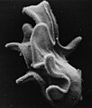 SEM bipinnaria.JPG 690 × 815; 65 KB
-
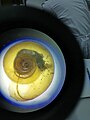 Shell under a microscope.jpg 3,120 × 4,160; 2.57 MB
-
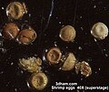 Shregg1a.jpg 450 × 378; 34 KB
-
 Sludge worm (265 31) Total preparation.jpg 3,751 × 2,400; 1.84 MB
-
 Squatinella rostrum - 400x - lateral view (19076204959).jpg 3,876 × 2,634; 6.85 MB
-
 Stratified Squamos Epithelium.png 757 × 670; 893 KB
-
 T-cell killing cancer cell.gif 300 × 300; 1.02 MB
-
 Tapeworm (26 2 14) Cross-section.jpg 2,773 × 1,775; 1.35 MB
-
 Tapeworm (265 06) Body segment.jpg 3,751 × 2,401; 1.8 MB
-
 Tapeworm (265 07) Body segment.jpg 3,751 × 2,401; 2.38 MB
-
 Tapeworm (265 15) Cross-section.jpg 3,751 × 2,400; 3.06 MB
-
 Tapeworm in love n2.JPG 3,840 × 2,160; 1.55 MB
-
 Tapeworm in love.JPG 3,840 × 2,160; 1.43 MB
-
 Tapeworm rostellum and hooks.jpg 1,080 × 1,920; 151 KB
-
 TardigradeEggsInShedCuticle.jpg 1,024 × 1,024; 255 KB
-
 The Biological bulletin (19757898313).jpg 1,998 × 2,332; 1.19 MB
-
 The Biological bulletin (19758011533).jpg 2,004 × 1,310; 608 KB
-
 Trematode (265 18) Dicrocoelium lanceolatum.jpg 3,750 × 2,400; 1.7 MB
-
 Trematode (265 32) Eggs in liver.jpg 3,750 × 2,400; 1.63 MB
-
 Trichinella spiralis in muscle tissue (265 16) In skeletal (striated) muscle tissue.jpg 3,750 × 2,400; 2.13 MB
-
 Tronco anisakis x100 pol cruzados.tif 2,080 × 1,544; 4.11 MB
-
 Two reniform nematodes.jpg 6,016 × 4,000; 8.17 MB
-
 UnknowLarvaVladivostok SEM.jpg 1,746 × 2,382; 470 KB
-
 Vaginalzyto-proestrus-ORS.jpg 4,000 × 3,000; 1.12 MB
-
 White worms (265 30) Total preparation.jpg 3,750 × 2,400; 1.67 MB
-
 Wrotki kolonijne.jpg 5,000 × 3,773; 6.11 MB
-
 Бабочка капустница (Pieris brassicae).jpg 7,009 × 5,493; 23.31 MB
-

-
 Долгоносик Sitona Sp.jpg 7,522 × 6,958; 26.63 MB
-
 Интерференционный узор крыла комара-звонца.tif 4,570 × 3,438; 108.9 MB
-
 Кишечник голотурии.jpg 1,492 × 995; 1.21 MB
-
 Комар - звонец.jpg 4,204 × 3,154; 18.03 MB
-
 Молодая медуза Aurelia aurita.jpg 1,517 × 1,565; 345 KB
-
 Молодая эфира Aurelia aurita.jpg 1,280 × 960; 137 KB
-
 Муха- горбатка.jpg 4,519 × 3,294; 7.87 MB
-
 Паук - волк крупным планом.tif 4,547 × 4,920; 117.43 MB
-
 Паук - волк.tif 4,592 × 3,448; 81.3 MB
-
 Раковина.png 2,592 × 1,944; 7.44 MB
-
 Спикулы (скелетные элементы) Iophon piceum (Porifera).png 1,105 × 1,419; 613 KB
-
 Спикулы Grayella pyrula (Porifera).png 957 × 1,283; 337 KB
-
 Тля, темное поле.jpg 6,165 × 4,466; 8.35 MB
-
 Трахейные пузыри личинки комара коретра. Темное поле + поляризация.jpg 4,152 × 3,136; 7.48 MB


.jpg)

_(20227843139).jpg)


_Cercaria_of_trematode.jpg)
.jpg)
.jpg)


_Cross-section_of_uterus_with_eggs.jpg)


_1D.JPG)


_Cross-section_of_leech_(Hirudinea).jpg)
_Cross-section_of_leech_(Hirudinea).jpg)

.jpg)
.jpg)
.jpg)
.jpg)
.jpg)
.jpg)
.jpg)
.jpg)


_From_bodies_of_Porifera,_sea_corals_and_holuthureans.jpg)

.png)

_Statoblast_of_Pectinatella_magnifica,_a_species_of_Bryozoa.jpg)


_Total_preparation.jpg)
.jpg)


_Cross-section.jpg)
_Body_segment.jpg)
_Body_segment.jpg)
_Cross-section.jpg)


.jpg)
.jpg)
_Dicrocoelium_lanceolatum.jpg)
_Eggs_in_liver.jpg)
_In_skeletal_(striated)_muscle_tissue.jpg)


_Total_preparation.jpg)

.jpg)
_%D0%B3%D1%83%D0%B1%D0%BA%D0%B8_Hymedesmia_similis_(Lundbeck,_1910).png)


_Iophon_piceum_(Porifera).png)
.png)

