Category:Microscopic images of unidentified plants
Jump to navigation
Jump to search
Subcategories
This category has the following 4 subcategories, out of 4 total.
Media in category "Microscopic images of unidentified plants"
The following 199 files are in this category, out of 199 total.
-
"То,что мы не видим".jpg 960 × 960; 365 KB
-
Analysing Plants Under a Microscope.jpg 2,560 × 1,536; 898 KB
-
Anatomia foliar.jpg 800 × 600; 83 KB
-
Art in science.jpg 548 × 556; 132 KB
-
Bifacial leaf cross section.jpg 678 × 800; 453 KB
-
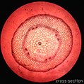
BIO cross section 2.jpg 1,276 × 1,284; 881 KB
-
BIO cross section 3.jpg 1,280 × 1,278; 899 KB
-
BIO cross section 4.jpg 1,258 × 1,304; 557 KB
-
Biological Boogie Man.jpg 1,280 × 960; 136 KB
-
Blattquerschnitt.jpg 1,280 × 1,024; 431 KB
-
Cells under a microscope.jpg 912 × 912; 181 KB
-
Chloroplasty.jpg 2,592 × 1,944; 439 KB
-
Clorofila 3.jpg 1,632 × 1,224; 844 KB
-
Corte anatômico.jpg 1,125 × 1,531; 1.2 MB
-
Coupe transversale d'une feuille de gymnosperme.jpg 1,536 × 2,048; 469 KB
-
Cuticle of leaf under microscope.JPG 2,447 × 2,447; 1.86 MB
-
Cuticle overlying upper epidermis in mesophyte leaf (35103215772).jpg 3,264 × 1,840; 1.23 MB
-
Cuticle overlying upper epidermis of mesophyte leaf (34686901852).jpg 3,264 × 1,840; 1.03 MB
-
Células vegetales (con azul de metileno).jpg 487 × 511; 79 KB
-
Drevesina.jpg 1,488 × 1,249; 1.49 MB
-
EB1911 Plants - transverse sections of leaves.jpg 830 × 472; 168 KB
-
Equine sorrel lower epidermis.jpg 1,944 × 2,592; 1.58 MB
-
Estoma microoscopio.jpg 1,200 × 1,600; 311 KB
-
Estoma.jpg 600 × 450; 21 KB
-
Estômatos.jpg 1,300 × 736; 393 KB
-
Flower petal (species unknown).png 741 × 830; 1.34 MB
-
Flower petal 1 400×.png 1,780 × 1,780; 3.23 MB
-
Flower petal 2 400×.png 1,690 × 1,690; 3.88 MB
-
Germination of pollen (255 27) Total preparation (pollen tube visible).jpg 3,748 × 2,399; 1.5 MB
-
Guard cells and stomata in succulent xerophyte leaf (35229487806).jpg 3,264 × 1,840; 957 KB
-
Guards cells in lower epidermis of mesophyte leaf (35103213882).jpg 3,264 × 1,840; 1.02 MB
-
Hoja al microscopio.jpg 653 × 607; 46 KB
-
Hydrophyte leaf (34357208213).jpg 3,264 × 1,840; 1.19 MB
-
Hydrophyte leaf (35228402066).jpg 2,767 × 1,559; 1.22 MB
-
Hydrophytic leaf micrograph.jpg 2,048 × 1,536; 2.69 MB
-
Inner Beauty of Plants.jpg 2,448 × 3,264; 1.52 MB
-
Iuoh;;.jpg 4,592 × 3,448; 3.84 MB
-
Lateral Root Origin (36223436805).jpg 3,264 × 1,840; 586 KB
-
Lateral Root Origin (36223438925).jpg 3,264 × 1,840; 820 KB
-
Lateral Root Origin (36223441325).jpg 3,264 × 1,840; 889 KB
-
Lateral Root Origin (36223443845).jpg 3,264 × 1,840; 853 KB
-
Leaf (255 14) Leaf semi-thick section.jpg 3,751 × 2,401; 1.77 MB
-
Leaf 123.jpg 1,176 × 999; 458 KB
-
Leaf cells - 02.jpg 2,592 × 3,872; 7.16 MB
-
Leaf cells - 03.jpg 3,872 × 2,592; 8.88 MB
-
Leaf cells with stomata.jpg 3,760 × 2,507; 8.2 MB
-
Leaf cells.jpg 3,000 × 2,008; 7.61 MB
-
Leaf epidermis 2.jpg 480 × 640; 301 KB
-
Leaf Epidermis.jpg 720 × 960; 84 KB
-
Leaf epithelium, guard cells, stomatal pore and peripheral chloroplasts.jpg 3,024 × 4,032; 962 KB
-
Leaf epithelium, stomata and guard cells.jpg 3,594 × 2,881; 2.34 MB
-
Leaf epithelium, stomata.jpg 2,762 × 2,012; 1.32 MB
-
Life teeming under the microscope.jpg 2,752 × 2,961; 708 KB
-
Longitudinal section of tree trunk. Wellcome M0010773.jpg 2,433 × 4,335; 2.96 MB
-
Lower foliari cell walls.jpg 601 × 453; 117 KB
-
Meiosis I.jpg 762 × 720; 131 KB
-
Meiosis Telophase I.jpg 1,280 × 720; 86 KB
-
Mesophyte leaf (34321748034).jpg 3,264 × 1,840; 1.05 MB
-
Mesophyte leaf (34321749754).jpg 2,609 × 1,470; 842 KB
-
Mesophyte leaf (34357209113).jpg 3,264 × 1,840; 1.12 MB
-
Mesophyte leaf (34810761446).jpg 3,264 × 1,840; 819 KB
-
Mesophyte x hydrophyte leaf (34381760130).jpg 3,264 × 1,840; 1.28 MB
-
Mesophytic leaf micrograph.jpg 2,048 × 1,536; 2.6 MB
-
Mia lab.jpg 1,600 × 1,200; 385 KB
-
Microscope plants 01.jpg 640 × 480; 74 KB
-
Microscope plants 02.jpg 640 × 480; 77 KB
-
Microscope plants 03.jpg 640 × 480; 58 KB
-
Microscope plants 04.jpg 640 × 480; 56 KB
-
Microscope plants 05.jpg 640 × 480; 78 KB
-
Microscope plants 06.jpg 640 × 480; 68 KB
-
Microscope plants 07.jpg 640 × 480; 59 KB
-
Microscope plants 08.jpg 640 × 480; 76 KB
-
Microscope plants 09.jpg 640 × 480; 62 KB
-
Microscope plants 10.jpg 640 × 480; 67 KB
-
Microscope plants 11.jpg 640 × 480; 88 KB
-
Microscope plants 12.jpg 640 × 480; 71 KB
-
Microscope plants 13.jpg 640 × 480; 57 KB
-
Microscope plants 14.jpg 640 × 480; 44 KB
-
Microscope plants 15.jpg 640 × 480; 111 KB
-
Microscope plants 16.jpg 640 × 480; 110 KB
-
Microscope plants 17.jpg 640 × 480; 88 KB
-
Microscope plants 18.jpg 640 × 480; 46 KB
-
Microscope plants 19.jpg 640 × 480; 90 KB
-
Microscope plants 20.jpg 640 × 480; 81 KB
-
Microscope plants 21.jpg 640 × 480; 100 KB
-
Microscopy image of a leaf.JPG 2,144 × 2,775; 555 KB
-
No Bacon, It is longitudinal section from stem of cotyledon.jpg 1,280 × 1,280; 108 KB
-
Open and Closed Stomata.jpg 3,024 × 4,032; 783 KB
-
Open stoma micrograph.tiff 1,280 × 1,100; 1.35 MB
-
Optical sectioning of pollen.jpg 1,521 × 1,950; 426 KB
-
Parenchyma.jpg 1,280 × 960; 83 KB
-
Parênquima aerífero.jpg 800 × 600; 155 KB
-
Phloem.jpg 715 × 750; 489 KB
-
Picture Natural History - No 314 315 - Section of Leaf.png 753 × 525; 701 KB
-
Picture Natural History - No 316 - Section of Leaf.png 499 × 364; 371 KB
-
Pitcher plant trichomes pollen.jpg 2,560 × 1,920; 2.36 MB
-
Plant cell type collenchyma.png 446 × 333; 295 KB
-
Plant cell type sclerenchyma fibers.png 448 × 335; 288 KB
-
Plant stem (250 26) Longitudal radial section - woody stem.jpg 3,752 × 2,401; 1.28 MB
-
Plant stem (250 27) Longitudal radial section - woody stem.jpg 3,751 × 2,400; 1.83 MB
-
Plant Under a Microscope.jpg 1,536 × 2,048; 532 KB
-
Plant under microscope.jpg 638 × 478; 73 KB
-
Root Apical Meristem cell.jpg 1,701 × 1,615; 526 KB
-
Root parenchyma (34877668995).jpg 3,264 × 1,840; 716 KB
-
Root tip.JPG 2,592 × 1,944; 1.26 MB
-
Rootmeristem100x1.jpg 1,024 × 768; 225 KB
-
Rootmeristem100x2.jpg 1,024 × 768; 295 KB
-
Rootmeristem400x1.jpg 1,024 × 768; 143 KB
-
Rootmeristem400x3.jpg 1,024 × 768; 231 KB
-
Rootmeristem400x4.jpg 1,024 × 768; 262 KB
-
Rootmeristem40x1.jpg 1,024 × 768; 190 KB
-
Rootmeristem40x2.jpg 1,024 × 768; 240 KB
-
Sclerids embedded in upper epidermis of hydrophyte leaf (35268633765).jpg 3,264 × 1,840; 1.25 MB
-
Sclerids in lower epidermis of hydrophyte leaf (35228410636).jpg 3,264 × 1,840; 1.27 MB
-
Sclerids in the hydrophyte leaf (35268634785).jpg 3,264 × 1,840; 1.09 MB
-
Sclerids in upper epidermis of hydrophyte (35228405986).jpg 3,264 × 1,840; 1.43 MB
-
Sclerids support in the lower epidermis of a hydrophyte leaf (35268636095).jpg 3,264 × 1,840; 1.22 MB
-
Scleroid (35228407326).jpg 3,264 × 1,840; 1.06 MB
-
Scleroid in epidermis of hydrophyte leaf (34810755436).jpg 3,264 × 1,840; 1.07 MB
-
Scleroid in lower epidermis of hydrophyte leaf (34686901272).jpg 3,264 × 1,840; 741 KB
-
Scleroid support in the hydrophyte leaf (35228408416).jpg 3,264 × 1,840; 1.37 MB
-
Seed Parachute.jpg 5,120 × 3,840; 7.14 MB
-
Stem Cross-Section.jpg 720 × 960; 50 KB
-
Stem of Dicotyledon C.S.png 781 × 752; 1.24 MB
-
Stem-Collenchyma100x4.jpg 1,024 × 768; 211 KB
-
Stem-Collenchyma100x5.jpg 1,024 × 768; 305 KB
-
Stem-Collenchyma100x6.jpg 1,024 × 768; 196 KB
-
Stem-Collenchyma400x3.jpg 1,024 × 768; 178 KB
-
Stem-Collenchyma40x1.jpg 1,024 × 768; 252 KB
-
Stem-Collenchyma40x2.jpg 1,024 × 768; 256 KB
-
Stem-Parenchyma100x1.jpg 1,024 × 768; 129 KB
-
Stem-Parenchyma400x1.jpg 1,024 × 768; 147 KB
-
Stem-Parenchyma400x6.jpg 1,024 × 768; 150 KB
-
Stem-Parenchyma400x8.jpg 1,024 × 768; 143 KB
-
Stem-Parenchyma40x1.jpg 1,024 × 768; 164 KB
-
Stem-Sclerenchvma100x2.jpg 1,024 × 768; 310 KB
-
Stoma and guard cells in succulent xerophyte leaf (34883081660).jpg 3,264 × 1,840; 1.01 MB
-
Stoma and guard cells in succulent xerophyte leaf (35229490736).jpg 3,264 × 1,840; 1.02 MB
-
Stoma.tif 640 × 550; 346 KB
-
Stomata and guard cells in upper epidermis of succulent (34810756046).jpg 3,264 × 1,840; 1.19 MB
-
Stomata in lower epidermis of mesophyte leaf (34634953011).jpg 3,264 × 1,840; 917 KB
-
Stomata observed on the rear side of a leaf.jpg 3,024 × 4,032; 1.45 MB
-
Stomata Pengo.jpg 1,600 × 1,200; 589 KB
-
Stomatal apparatus.jpg 738 × 690; 114 KB
-
Succulent xerophyte leaf (34426136954).jpg 3,264 × 1,840; 1.53 MB
-
Succulent xerophyte leaf (35229483216).jpg 3,264 × 1,840; 1.1 MB
-
T.S. of monocot stem.jpg 727 × 696; 128 KB
-
The pollen and the wing.jpg 1,032 × 774; 186 KB
-
The Skull within.jpg 2,448 × 3,264; 1.51 MB
-
Trachid and vessels within xylem bundle of mesophyte leaf (34605001012).jpg 3,264 × 1,840; 1.12 MB
-
Transverse section of collenchyma.jpg 1,280 × 960; 76 KB
-
Transverse section of phloem.jpg 960 × 1,280; 102 KB
-
Transverse section of xylem.jpg 960 × 1,280; 98 KB
-
Tubos cribosos.jpg 2,824 × 2,600; 1.26 MB
-
Vascular bundle in hydrophyte leaf (33958476013).jpg 3,264 × 1,840; 1.16 MB
-
View Inside Microscope.jpg 2,513 × 2,513; 1.07 MB
-
Violet darkfield.jpg 3,872 × 2,592; 5.04 MB
-
Violet glandular trichomes.jpg 700 × 1,050; 606 KB
-
Xerophyte leaf (34321742324).jpg 3,264 × 1,840; 1.07 MB
-
Xerophyte leaf (34686910152).jpg 3,264 × 1,840; 1.08 MB
-
Xerophyte leaf Succulent (35229491476).jpg 3,264 × 1,840; 1.07 MB
-
Xerophyte leaf x mesophyte leaf (34381760970).jpg 3,264 × 1,840; 1.44 MB
-
Xerophyte leaf x Mesophyte leaf (34810763066).jpg 3,264 × 1,840; 1.36 MB
-
Xerophytic Leaf Cross Section Stained Microscope Slide.jpg 1,280 × 960; 307 KB
-
Xerophytic leaf micrograph.jpg 2,048 × 1,536; 2.8 MB
-
Xilema em corte longitudinal.jpg 1,600 × 1,200; 446 KB
-
Xylem and phloem tissue.jpg 720 × 712; 112 KB
-
Xylem.jpg 2,048 × 1,536; 1.05 MB
-
Ευκαρυωτικά κύτταρα από μικροσκόπιο.JPG 4,000 × 3,000; 5.29 MB
-
Ευκαρυωτικά κύτταρα σε μικροσκόπιο.JPG 4,000 × 3,000; 5.59 MB
-
Водоросли водная сеточка, темное поле.jpg 3,811 × 2,862; 8.36 MB
-
Волоски на поверхности листа.tif 1,024 × 943; 2.77 MB
-
Деплазмолиз клетки.jpeg 2,448 × 3,264; 1.05 MB
-
Капля эфирного масла.png 640 × 480; 579 KB
-
Клетки водоросли с хлоропластами.jpg 4,063 × 3,050; 3.44 MB
-
Клетки мха в поляризованном свете.jpg 4,253 × 3,157; 13.56 MB
-
Клетки мха.jpg 4,072 × 3,064; 9.24 MB
-
Коетка кое чего.jpg 1,920 × 2,560; 2.16 MB
-
Микрофотография такни зоны всасывания.jpg 1,218 × 1,624; 151 KB
-
Окаймлённая пора ( похожа на глаз дракона).jpg 963 × 691; 388 KB
-
Поперечное сечение сеточки сивы в поляризованном свете.jpg 6,678 × 7,154; 29.17 MB
-
Пыльник.jpg 4,912 × 3,264; 2.16 MB
-
Сорус спорофита папоротника.jpg 1,224 × 1,056; 366 KB
-
Спорангій папороті.jpg 475 × 437; 119 KB
-
Срез иголки сосны.jpg 1,600 × 1,200; 730 KB
-
Срез растения в ультрафиолете.jpg 4,608 × 3,456; 7.7 MB
-
Стерильные пыльцевые зерна тетраплоидной формы яблони.jpg 1,152 × 1,046; 247 KB
-
Строение стебля двудольного растения.jpg 1,792 × 1,344; 342 KB
-
Строение эпителия листа.JPG 2,204 × 3,920; 1.15 MB
-
То, что незаметно глазу.jpg 810 × 1,080; 181 KB
-
Устьица на листе.jpg 652 × 615; 57 KB
-
Устьице листа хойи под микроскопом.jpg 780 × 1,040; 120 KB
-
Флуоресценция листа под микроскопом.JPG 4,912 × 3,264; 2.03 MB
-
Хлоропласты 40.png 2,592 × 1,944; 8.75 MB
-
Хлоропласты 400.png 2,592 × 1,944; 6.44 MB
-
Чаинки.jpg 2,560 × 1,707; 1.38 MB
