Category:Microscopic images of human cells
Jump to navigation
Jump to search
This category is still missing many files and subcategories.
Media in category "Microscopic images of human cells"
The following 47 files are in this category, out of 47 total.
-
 38F3-ChkNFH-DAPI-Shsy5y.jpg 1,315 × 1,033; 493 KB
-
-
-
-
 Cel interst Cajal Estructura.png 515 × 536; 203 KB
-
 Cel interst Cajal Prolongaciones.png 527 × 542; 289 KB
-
 Celulas mucosa bucal humana.jpg 640 × 521; 81 KB
-
 Co-transfected HPCA-mTFP1 and EYFP-Mem HEK 293 cells.png 1,024 × 1,024; 727 KB
-
 Cèl·lules de la mucosa bucal 20180407 113911.jpg 4,128 × 3,096; 653 KB
-
 Cèl·lules de la mucosa bucal 20180407 113914.jpg 4,128 × 3,096; 675 KB
-
 Cèl·lules de la mucosa bucal 20180407 114212.jpg 4,128 × 3,096; 595 KB
-
 Endoteliales Cultivo.png 697 × 722; 503 KB
-
 Endoteliocitos SEM.png 842 × 792; 431 KB
-
 Epithelial Tissues Pseudostratified Columnar Epithelium (26915090687).jpg 3,264 × 1,840; 1.41 MB
-
 Epithelial Tissues Pseudostratified Columnar Epithelium (26915091897).jpg 3,264 × 1,840; 817 KB
-
 Epithelial Tissues Pseudostratified Columnar Epithelium (40883906325).jpg 3,264 × 1,840; 1.3 MB
-
 Epithelial Tissues Pseudostratified Columnar Epithelium (41783110841).jpg 3,264 × 1,840; 1.29 MB
-
 Epithelial Tissues Pseudostratified Columnar Epithelium (41783111911).jpg 3,264 × 1,840; 941 KB
-

-
 Epithelial Tissues Simple Cuboidal Epithelium (39914417240).jpg 3,264 × 1,840; 1.53 MB
-
 Epithelial Tissues Simple Cuboidal Epithelium (39914418360).jpg 3,264 × 1,840; 1.38 MB
-
 Epithelial Tissues Simple Cuboidal Epithelium (39914419200).jpg 3,264 × 1,840; 4.91 MB
-
 Epithelial Tissues Simple Cuboidal Epithelium (39914419940).jpg 3,264 × 1,840; 4.89 MB
-
 Epithelial Tissues Simple Cuboidal Epithelium (41681552432).jpg 3,264 × 1,840; 7.31 MB
-
 Epithelial Tissues Simple Cuboidal Epithelium (41681552782).jpg 3,264 × 1,840; 902 KB
-
 Epithelial Tissues Simple Cuboidal Epithelium (41721682831).jpg 3,264 × 1,840; 1.44 MB
-
 Fagocytoza.jpg 2,560 × 1,440; 357 KB
-
 Fluo-4 loaded HEK 293 cells.png 1,024 × 1,024; 1.81 MB
-
 Human Cells (6842745203).jpg 1,050 × 844; 407 KB
-
 Human cheek cell.jpg 2,337 × 2,313; 1.36 MB
-
 Human Cheek Cells (Iodine Stain).jpg 1,669 × 1,734; 392 KB
-
 Human Cheek Cells (Methylene Blue Stain).jpg 1,198 × 1,239; 158 KB
-
 Human pseudostratified ciliated columnar epithelium 13.png 1,400 × 1,295; 2.48 MB
-
 Human umbilical vein endothelial cells.jpg 2,659 × 2,618; 1.29 MB
-
 Human-meniscus-cells-gruber-et-al-2008.tiff 1,300 × 1,212; 1.86 MB
-
 MeT5a.png 1,024 × 765; 1.52 MB
-
 Mouth cells.jpg 400 × 300; 74 KB
-
 Neurofilament staining of human cerebellum.jpg 842 × 550; 100 KB
-
 Nucleus in human cheek cell 1.jpg 640 × 480; 187 KB
-
 Nucleus in human cheek cell 2.jpg 640 × 480; 187 KB
-
 Nucleus in human cheek cell.jpg 640 × 480; 185 KB
-
 Nucleus in human cheek cells 3.jpg 640 × 480; 193 KB
-
 Nucleus in human cheek cells 4.jpg 640 × 480; 204 KB
-
 Nucleus in human cheek cells.jpg 640 × 480; 205 KB
-
 SK8-18-2 human derived cells, fluorescence microscopy (29942101073).jpg 2,758 × 2,214; 450 KB
-
 Spreadthelove - Treated human melanoma cell A375.jpg 376 × 336; 50 KB
-
 Spreadthelove - Untreated human melanoma cell A375.jpg 244 × 314; 18 KB

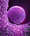
_virus_particles_(teal_color).jpg)
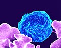
_infected_with_malaria_parasites_(blue)_with_the_small_bumps_on_the_infected_cell_show_how_the_parasite_remodels_its_host_cell_by_forming_protrusions_called_%27knobs%27_on_the_surface_(2).jpg)
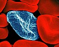
_infected_with_malaria_parasites_(blue)_with_the_small_bumps_on_the_infected_cell_show_how_the_parasite_remodels_its_host_cell_by_forming_protrusions_called_%27knobs%27_on_the_surface.jpg)


.jpg)
.jpg)
.jpg)
.jpg)
.jpg)
_(cropped).jpg)
.jpg)
.jpg)
.jpg)
.jpg)
.jpg)
.jpg)
.jpg)


.jpg)

.jpg)
.jpg)


.jpg)

