Category:Microscopic images of cell nucleus stained with DAPI in HeLa cells
Jump to navigation
Jump to search
Media in category "Microscopic images of cell nucleus stained with DAPI in HeLa cells"
The following 15 files are in this category, out of 15 total.
-
-
-
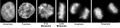
 Confocal micrograph of stained HeLa.jpg 1,345 × 1,345; 883 KB
-
 Cuerpos de Cajal.jpg 600 × 450; 259 KB
-
 HELA cells stained with DAPI and Phalloidin.tiff 1,376 × 1,038; 4.1 MB
-
-
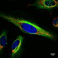
 Ki67-Tubulin-2.jpg 1,156 × 874; 542 KB
-
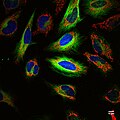
 Ki67-Tubulin.jpg 1,186 × 870; 743 KB
-
 Microtúbulos. Inmuno histoquímica Tubulinas alfa y gama.TIF 1,457 × 1,287; 2.06 MB
-
 Mitochondria in living HeLa cells.jpg 719 × 607; 27 KB
-
 Mitosepanel es.jpg 1,050 × 246; 27 KB
-
 Mitosepanel-rus.tif 941 × 234; 234 KB
-
 Mitosepanel.jpg 1,050 × 246; 39 KB
-
 Multicolor fluorescence image of a living HeLa cell.jpg 1,024 × 1,024; 148 KB
-
 Multicolor fluorescence image of living HeLa cells.jpg 1,024 × 1,024; 184 KB


