Category:Magnetic resonance imaging of the normal brain
Jump to navigation
Jump to search
Please, make sure the normality of the images
Media in category "Magnetic resonance imaging of the normal brain"
The following 133 files are in this category, out of 133 total.
-
 26638.medium-emphasizing-corpus-callosum.png 455 × 460; 202 KB
-
 26638.medium.jpg 455 × 460; 75 KB
-
 3D VR Image 200.jpg 201 × 202; 36 KB
-
 3d wrap-around artifact head.jpg 405 × 455; 79 KB
-
 3Dbrain.gif 256 × 256; 584 KB
-
 3DPX-002386 Atrophic Brain Nevit Dilmen.stl 5,120 × 2,880; 14.31 MB
-
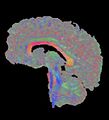
 3DPX-003145 Fractional Anisotrpy Tractography White Matter NevitDilmen.stl 5,120 × 2,880; 5.43 MB
-
 3DPX-003765 3DModel of Brain Nevit Dilmen.stl 5,120 × 2,880; 3.93 MB
-
 AGF ArctanLR.jpg 720 × 539; 145 KB
-
 Air-Ventriculograph CT MRI film.jpg 1,800 × 2,400; 510 KB
-
 Aktivitaethinten.jpg 220 × 239; 24 KB
-
 Amgydala.jpg 1,181 × 1,357; 223 KB
-
 AnisoColor.tif 1,112 × 600; 1.91 MB
-
 Atlanto-occipital joint.jpg 933 × 619; 54 KB
-
 MRI EGC sagittal.png 576 × 576; 306 KB
-
 Brain chrischan 300.gif 300 × 300; 756 KB
-
 Brain chrischan 600.gif 600 × 600; 2.56 MB
-
 Brain chrischan thalamus.jpg 860 × 860; 71 KB
-
 Brain Diff 120045 rgbca cr.png 469 × 513; 498 KB
-
 Brain MRI 103223 rgbca.png 474 × 498; 535 KB
-
 Brain MRI 103337 rgbca.png 528 × 528; 446 KB
-
 Brain MRI 103618 rgbca.png 500 × 541; 594 KB
-
 Brain MRI 110439 rgbca.png 481 × 537; 605 KB
-
 Brain MRI 112010 rgbca.png 398 × 504; 489 KB
-
 Brain MRI 112445 rgbca.png 438 × 486; 491 KB
-
 Brain MRI 115042 rgbc pd t2 diff.png 474 × 520; 549 KB
-
 Brain MRI 120329 T1 Sag 82M.png 552 × 510; 197 KB
-
 Brain MRI 123515 rgbca.png 507 × 586; 384 KB
-
 Brain MRI 124054 rgbca.png 504 × 561; 539 KB
-
 Brain MRI 131058 rgbca.png 515 × 588; 540 KB
-
 Brain MRI 131444.png 448 × 512; 192 KB
-
 Brain MRI 131666.png 480 × 512; 183 KB
-
 Brain MRI 133204 rgbca.png 420 × 450; 362 KB
-
 Brain MRI 133405 rgbca.png 478 × 562; 458 KB
-
 Brain MRI 135119 rgbca.png 459 × 507; 491 KB
-
 Brain MRI 142044 rgbca.png 408 × 468; 399 KB
-
 Brain MRI 145729 t1 pd t2 18F Epil.png 482 × 554; 517 KB
-
 Brain MRI 150443 rgbca t1 t2 t2STIR misreg.png 419 × 522; 493 KB
-
 Brain MRI 4yF 133316-rgbca.png 420 × 463; 365 KB
-
 Brain MRI FLAIR Cor 142219.png 319 × 426; 92 KB
-
 Brain Mri nevit.svg 579 × 778; 565 KB
-
 Brain MRI t2 142301.png 440 × 448; 168 KB
-
 Brain MRI.gif 165 × 186; 207 KB
-
 Brain regions on T1 MRI.png 933 × 717; 763 KB
-
 Caudate Nucleus Structural MRI.png 577 × 769; 358 KB
-
 Caudate nucleus.jpg 1,181 × 1,419; 323 KB
-
 Cavum septi pellucidi - Cavum vergae.jpg 853 × 992; 135 KB
-
 Cavum septi pellucidi fetal.jpg 802 × 1,161; 145 KB
-
 Cavum septi pellucidi MRT cor T1.jpg 1,180 × 1,087; 133 KB
-
 Choclea and vestibular system 160009.jpg 2,560 × 1,920; 1.79 MB
-
 CO2-O2-fMRI-all-over-time.png 2,800 × 4,269; 5.34 MB
-
 Cochlea and semicirculars MRI 115700 MAIP.png 490 × 506; 263 KB
-
 Cochlea and vestibular system.gif 480 × 480; 4.71 MB
-
 3DTomo.jpg 409 × 544; 59 KB
-
 Corps calleux Sagittal.JPG 479 × 498; 32 KB
-
 Corpuis callosum.png 624 × 624; 302 KB
-
 CPM3.jpg 722 × 903; 67 KB
-
 Cuneus.png 661 × 550; 243 KB
-
 Diffusion 105025 rgbc2.png 522 × 534; 242 KB
-
 Diffusion MRI 105025 rgbc.png 522 × 534; 246 KB
-
 DTI Brain Tractographic Image A panal.jpg 692 × 575; 243 KB
-
 DTI Brain Tractographic Image Set.jpg 1,500 × 1,248; 921 KB
-
 DTI-sagittal-fibers.jpg 1,021 × 952; 294 KB
-
 DTI-sagittal-xyzrgb.jpg 1,021 × 1,125; 64 KB
-
 Faisceau pedicule.jpg 600 × 600; 48 KB
-
 FastFission-brain-bild52.png 178 × 227; 8 KB
-
 Fiducials-am-hi-erc.png 1,443 × 642; 1.17 MB
-
 FMRI Brain Scan.jpg 287 × 234; 16 KB
-
 Fmrtuebersicht.jpg 1,025 × 915; 160 KB
-
 Functional magnetic resonance imaging.jpg 250 × 208; 11 KB
-
 GarpenBrain.jpg 1,280 × 1,024; 677 KB
-
 Generating Magnetic Resonance Spectroscopy Imaging Data of Brain Tumours from Linear, Non-Linear and Deep Learning Models.pdf 1,275 × 1,650, 9 pages; 1.25 MB
-
 Globus Pallidus structural MRI.png 577 × 769; 357 KB
-
Head MRI, sagittal plane, T₂ weighted.webm 4.9 s, 512 × 512; 1.5 MB
-
 High Resolution FMRI of the Human Brain.gif 512 × 256; 1.06 MB
-
 Hippocampus-mri.jpg 510 × 510; 71 KB
-
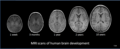 Human brain development 1wk-3mo-1yr-2yr-10yr-T1W-MRI.PNG 1,023 × 414; 186 KB
-
 Human cerebral cortex.png 411 × 384; 84 KB
-
 Hypothalamus.jpg 236 × 248; 11 KB
-
 Inner ear Chochlea and vestibular system 115700 MAIP3.png 494 × 468; 208 KB
-
 Labeledbrain.jpg 860 × 860; 78 KB
-
 Local field 400.jpg 402 × 212; 14 KB
-
 Max contrast Brain MRI 131058 rgbcb.png 515 × 588; 417 KB
-
 Max contrast Brain MRI 131058 rgbce.png 498 × 569; 666 KB
-
-
-
 Mni-autoreg-00-model.png 362 × 435; 83 KB
-
 MR bakke-naese.gif 384 × 384; 3.54 MB
-
 Mr cranial sinusvene.jpg 860 × 928; 84 KB
-
 MR side.gif 216 × 256; 943 KB
-
 Mra-mip.jpg 450 × 527; 101 KB
-
 MRI 123900.png 461 × 542; 580 KB
-
 Mri brain side view-emphasizing-corpus-callosum.png 600 × 600; 1.38 MB
-
 Mri brain side view.jpg 900 × 900; 225 KB
-
 MRI brain.jpg 238 × 253; 14 KB
-
 Mri head 3dani 1 small bionerd.gif 200 × 243; 734 KB
-
 MRI head side.jpg 256 × 256; 10 KB
-
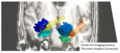
 MRI Location Amygdala up.png 704 × 666; 252 KB
-
 MRI Location Hippocampus up..png 667 × 649; 289 KB
-
 MRI T2 Brain axial image.jpg 900 × 1,119; 390 KB
-
 MRI.png 1,512 × 1,056; 6.77 MB
-
 Mrt big.jpg 820 × 754; 192 KB
-
 Murphy 2013 brain MRE complete.tif 2,049 × 1,633; 1.82 MB
-
 Murphy 2013 brain MRE reduced.png 780 × 1,626; 370 KB
-
 Murphy 2013 brain MRE with wave image.png 1,414 × 1,626; 700 KB
-
 Nucleus accumbens sag.jpg 846 × 844; 88 KB
-
 Ofc1.jpg 629 × 600; 57 KB
-
 Pallidus.jpg 1,181 × 1,428; 320 KB
-
 Pathways of the dorsal and ventral.png 567 × 603; 261 KB
-
 PET-IRM-cabeza-Keosys.JPG 1,280 × 1,024; 117 KB
-
 PLoS ONE 0000222 g001 MRS.jpg 2,048 × 717; 196 KB
-
 Powell2004Fig1A.jpeg 392 × 486; 35 KB
-
 Powell2004Fig1A.png 326 × 206; 81 KB
-
 Precuneus.png 699 × 700; 251 KB
-
 Putamen structuralMRI.png 577 × 769; 358 KB
-
 Putamen.jpg 1,181 × 1,423; 324 KB
-
 RetroOrbita 124440 MRI FLAIR.png 412 × 473; 123 KB
-
 Sassdasda.JPG 213 × 239; 15 KB
-
Stereoscopic-three-dimensional-visualization-applied-to-multimodal-brain-images-clinical-Video3.ogv 12 s, 946 × 573; 4.11 MB
-
 Striatum Structural MRI.png 577 × 769; 359 KB
-
Structural MRI animation.ogv 5.6 s, 256 × 256; 1.76 MB
-
 Substantia Nigra.jpg 849 × 846; 99 KB
-
 SWI 4Tesla.png 269 × 293; 88 KB
-
 T1t2PD.jpg 541 × 239; 30 KB
-
 Thalamus.jpg 236 × 248; 11 KB
-
 Thalamus2008.jpg 1,181 × 1,402; 327 KB
-
 TORNAI-SpectrumOfMedicalImaging.jpg 720 × 504; 117 KB
-
 U fibres big.JPG 1,638 × 1,458; 185 KB
-
 User-FastFission-brain.gif 213 × 231; 444 KB
-
 Ventral midbrain.png 994 × 1,017; 922 KB
-
 Vet brain 123501-rgbca.png 490 × 456; 534 KB
-
 Webysther 635864498758569254 - Imagem por ressonância magnética.png 480 × 480; 170 KB
-
 Магнитно-резонансная томография.jpg 960 × 1,280; 97 KB


