Category:Lateral ventricles
Jump to navigation
Jump to search
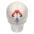
cavity in each of the hemispheres of the human brain  Representació 3D del sistema ventricular de l'encèfal humà. Els ventricles laterals són en vermell. | |||||
| Upload media | |||||
| Instance of |
| ||||
|---|---|---|---|---|---|
| Subclass of |
| ||||
| Part of | |||||
| |||||
Subcategories
This category has the following 5 subcategories, out of 5 total.
Media in category "Lateral ventricles"
The following 116 files are in this category, out of 116 total.
-
 26638.medium-emphasizing-corpus-callosum.png 455 × 460; 202 KB
-
 26638.medium.jpg 455 × 460; 75 KB
-
 3DPX-003027 Lateral ventricles MRI NevitDilmen.stl 5,120 × 2,880; 1.71 MB
-
 3DPX-003132 Cast of Lateral ventricles in Hydrocephaly NevitDilmen.stl 5,120 × 2,880; 6.43 MB
-
A-Model-of-Ischemia-Induced-Neuroblast-Activation-in-the-Adult-Subventricular-Zone-pone.0005278.s009.ogv 5.2 s, 976 × 816; 2.77 MB
-
-
-
 Alzheimer's disease brain preclinical.jpg 2,778 × 2,208; 3.91 MB
-
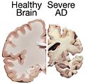
 Alzheimer's disease brain severe.jpg 3,000 × 2,250; 3.86 MB
-
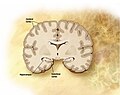
 Alzheimers brain.jpg 500 × 492; 31 KB
-
 Areas of Lateral ventricle - 01.png 1,200 × 1,200; 973 KB
-
 Areas of Lateral ventricle - 02.png 1,200 × 1,200; 1.12 MB
-
 Areas of Lateral ventricle - 03.png 1,200 × 1,200; 962 KB
-
 Areas of Lateral ventricle - 04.png 1,200 × 1,200; 1.12 MB
-
 Areas of Lateral ventricle - 05.png 1,200 × 1,200; 972 KB
-
 Areas of Lateral ventricle - 06.png 1,200 × 1,200; 1,000 KB
-
 Areas of Lateral ventricle - animation.gif 600 × 600; 6.03 MB
-
 Areas of Lateral ventricle -- 01.png 1,200 × 1,200; 264 KB
-
 Areas of Lateral ventricle -- 02.png 1,200 × 1,200; 311 KB
-
 Areas of Lateral ventricle -- 03.png 1,200 × 1,200; 270 KB
-
 Areas of Lateral ventricle -- 04.png 1,200 × 1,200; 304 KB
-
 Areas of Lateral ventricle -- 05.png 1,200 × 1,200; 326 KB
-
 Areas of Lateral ventricle -- 06.png 1,200 × 1,200; 363 KB
-
 Areas of Lateral ventricle -- animation.gif 600 × 600; 2.3 MB
-
 Basal ganglia 1.jpg 960 × 720; 89 KB
-
 Bourgery & Jacob-cf16.jpg 1,785 × 2,574; 2.53 MB
-

-
 Brain abscess simple brain CT.jpg 1,200 × 1,242; 138 KB
-
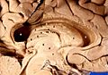
 Brain dissected after Prof. Nicolas method 2.jpg 1,500 × 1,125; 172 KB
-
 Brain dissected after Prof. Nicolas method.jpg 1,500 × 1,125; 171 KB
-
 Brain herniation types-2.svg 619 × 684; 415 KB
-
 Brain-Negro-Gorilla.png 400 × 335; 93 KB
-
 Cavum.jpg 1,660 × 683; 290 KB
-

-
 Formalin-fixated human brain1.jpg 263 × 329; 128 KB
-
 Formalin-fixated human brain3.jpg 263 × 280; 94 KB
-
 Gray664-es.png 1,000 × 740; 464 KB
-
 Gray717.png 500 × 568; 102 KB
-
 Gray718.png 550 × 590; 115 KB
-
 Gray734.png 500 × 370; 80 KB
-
 Gray735.png 450 × 451; 33 KB
-
 Gray736.png 450 × 332; 20 KB
-
 Gray737.png 500 × 589; 105 KB
-
 Gray738.png 450 × 376; 32 KB
-
 Gray739.png 500 × 379; 186 KB
-
 Gray742.png 500 × 572; 57 KB
-
 Gray743.png 450 × 542; 45 KB
-
 Gray748.png 500 × 510; 66 KB
-
 Gray749.png 500 × 308; 22 KB
-
 Gray750.png 417 × 516; 79 KB
-
 Hirnventrikel.gif 640 × 640; 6.82 MB
-
 Human brain frontal (coronal) section.JPG 702 × 487; 41 KB
-
 Human brain inferior-medial view description 2.JPG 702 × 491; 68 KB
-
 Human brain inferior-medial view description 3.JPG 702 × 491; 63 KB
-
 Human brain inferior-medial view.JPG 702 × 491; 52 KB
-
 Human brain left dissected midsagittal view description 2.JPG 701 × 488; 50 KB
-
 Human brain left dissected midsagittal view description.JPG 701 × 488; 50 KB
-
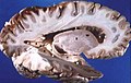
 Human brain left dissected midsagittal view.JPG 701 × 488; 46 KB
-
 Human brain right dissected lateral view description.JPG 653 × 413; 40 KB
-
 Human brain right dissected lateral view.JPG 653 × 413; 37 KB
-
 Huntington.jpg 722 × 902; 80 KB
-
Identification-of-the-Rostral-Migratory-Stream-in-the-Canine-and-Feline-Brain-pone.0036016.s001.ogv 9.0 s, 960 × 540; 795 KB
-
 INFARCT.jpg 327 × 409; 90 KB
-
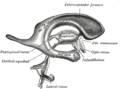
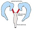 Interventricularforamina.jpg 351 × 295; 33 KB
-
 Intracerebral hemorrhage.jpg 400 × 330; 41 KB
-
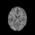 Kasraie scalped 3d010r cal BET2 0001.png 405 × 405; 61 KB
-
 Lateral ventricle - 01.png 1,200 × 1,200; 1,005 KB
-
 Lateral ventricle - 02.png 1,200 × 1,200; 1.17 MB
-
 Lateral ventricle - 03.png 1,200 × 1,200; 998 KB
-
 Lateral ventricle - 04.png 1,200 × 1,200; 1.17 MB
-
 Lateral ventricle - 05.png 1,200 × 1,200; 994 KB
-
 Lateral ventricle - 06.png 1,200 × 1,200; 1,010 KB
-
 Lateral ventricle - animation.gif 600 × 600; 7.04 MB
-
 Lateral ventricle -- 01.png 600 × 600; 100 KB
-
 Lateral ventricle -- 02.png 600 × 600; 112 KB
-
 Lateral ventricle -- 03.png 600 × 600; 102 KB
-
 Lateral ventricle -- 04.png 600 × 600; 111 KB
-
 Lateral ventricle -- 05.png 600 × 600; 123 KB
-
 Lateral ventricle -- 06.png 600 × 600; 138 KB
-
 Lateral ventricle -- animation.gif 600 × 600; 2.92 MB
-
 Lateral ventricle small.gif 200 × 200; 549 KB
-
 Lateral ventricle.gif 600 × 600; 3.95 MB
-
 Lateral ventricle.png 800 × 455; 315 KB
-
 Lateral Ventricles - DK ATLAS.png 1,200 × 900; 515 KB
-
Lateral Ventricles Demonstration by Dr Sanjoy Sanyal - MUA Neuroscience Lab 1 of 2.webm 7 min 33 s, 352 × 288; 54.74 MB
-
Lateral Ventricles Demonstration by Dr Sanjoy Sanyal - MUA Neuroscience Lab 2 of 2.webm 6 min 2 s, 352 × 288; 47.09 MB
-
 Lateral ventricles.jpg 960 × 720; 127 KB
-
 Lawrence 1960 2.32.png 2,000 × 1,088; 537 KB
-
 Lawrence 1960 2.33.png 1,504 × 1,460; 692 KB
-
 Lawrence 1960 2.34.png 1,632 × 1,152; 461 KB
-
 Lawrence 1960 2.35.png 2,260 × 1,628; 1,024 KB
-
 Mesial Temporal Sclerosis.jpg 296 × 320; 24 KB
-
 MRI brain tumor.jpg 1,839 × 1,737; 2.14 MB
-
 MRI of brain with sub-ependymal giant cell astrocytoma.jpg 387 × 512; 121 KB
-
 Pvx8133.tmp.jpg 339 × 297; 18 KB
-
 Sega gross.jpg 717 × 512; 221 KB
-
 Sheep Brain Dissection 3.jpg 1,600 × 1,200; 389 KB
-
 Slide10gg.JPG 960 × 720; 88 KB
-
 Slide11qq.JPG 960 × 720; 89 KB
-
 Slide1ff.JPG 960 × 720; 86 KB
-
 Slide1gg.JPG 960 × 720; 88 KB
-
 Slide2GRE.JPG 960 × 720; 83 KB
-
 Slide2oo.JPG 960 × 720; 66 KB
-
 Slide3GRE.JPG 960 × 720; 79 KB
-
 Slide3oo.JPG 960 × 720; 71 KB
-
 Slide4oo.JPG 960 × 720; 89 KB
-
 Slide5oo.JPG 960 × 720; 83 KB
-
 Slide7ff.JPG 960 × 720; 85 KB
-
 Sobo 1909 637.png 1,061 × 1,050; 3.19 MB
-
 Sobo 1909 639.png 564 × 806; 1.74 MB
-
 Space Occupying Lesion with Metastatic Infiltration.jpg 1,504 × 1,737; 433 KB
-
-
-
-
 The surgical approaches to the lateral ventricle.png 1,524 × 1,128; 1.6 MB
-
 Vesalius 609c.png 923 × 736; 400 KB


_--_showing_a_small_ring-enhancing_lesion_with_mild_surrounding_edema_adjacent_to_the_ventricular_catheter_and_ventricular_dilatation..jpg)


_section.JPG)


