Category:Ixodidae anatomy
Jump to navigation
Jump to search
Media in category "Ixodidae anatomy"
The following 27 files are in this category, out of 27 total.
-
 1902 Cattle ticks (Ixodoidea) of the United States p467 Fig. 227.jpg 1,812 × 898; 587 KB
-
 Acari, Myriopoda et Scorpiones hucusque in Italia reperta (1892) (16562274027).jpg 2,428 × 4,020; 1,013 KB
-
 Cattle ticks (Ixodoidea) of the United States (1902) (20398744510).jpg 2,631 × 4,359; 1.03 MB
-
 Cattle ticks (Ixodoidea) of the United States (1902) (20577791372).jpg 2,631 × 4,359; 1.1 MB
-
 Cattle ticks (Ixodoidea) of the United States (1902) (20586730455).jpg 2,476 × 4,222; 1.74 MB
-
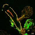
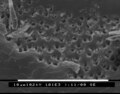 Elektronen microscopische foto teek oog.tif 1,424 × 1,120; 1.52 MB
-
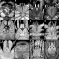 Haug 2020 Feeding apparatuses of different extant representatives of Arachnida.png 1,625 × 1,625; 1.7 MB
-
 Ixodholfemale.gif 342 × 390; 10 KB
-
 Ixodholmale.gif 335 × 389; 8 KB
-
 Ixodid tick structure.jpg 4,941 × 2,445; 2.06 MB
-
 JCD1 Kapfer A10 1.jpg 768 × 576; 100 KB
-

-

-

-

-

-

-

-

-
 Rhipicephalus annulatus claw.png 327 × 455; 46 KB
-
 Snuit van teek 2.jpg 352 × 288; 16 KB
-
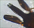 Snuit van teek.jpg 352 × 288; 12 KB
-
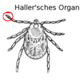 Tick (PSF) Hallersches Organ Ort.png 356 × 351; 28 KB
-
 Tick Salivary Gland Infected with Langat Virus (47974625372).jpg 2,048 × 2,048; 4.07 MB
-
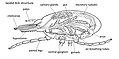
 The ventral aspect of a tick. Pen and ink drawing by A.J.E. Wellcome V0022611.jpg 2,300 × 3,090; 2.98 MB
-
 Колюще-сосущий тип ротового аппарата иксодового клеща.jpg 1,300 × 978; 1.64 MB
-
 Хоботок клеща.jpg 960 × 720; 28 KB
_of_the_United_States_p467_Fig._227.jpg)
_(16562274027).jpg)
_of_the_United_States_(1902)_(20398744510).jpg)
_of_the_United_States_(1902)_(20577791372).jpg)
_of_the_United_States_(1902)_(20586730455).jpg)


_-_Mites_-_Collection_Anthonie_Cornelis_Oudemans.jpeg)
_-_Mites_-_Collection_Anthonie_Cornelis_Oudemans.jpeg)
_-_Mites_-_Collection_Anthonie_Cornelis_Oudemans.jpeg)
_-_Mites_-_Collection_Anthonie_Cornelis_Oudemans.jpeg)
_-_Mites_-_Collection_Anthonie_Cornelis_Oudemans.jpeg)
_-_Mites_-_Collection_Anthonie_Cornelis_Oudemans.jpeg)
_-_Mites_-_Collection_Anthonie_Cornelis_Oudemans.jpeg)
_-_Mites_-_Collection_Anthonie_Cornelis_Oudemans.jpeg)


_Hallersches_Organ_Ort.png)
.jpg)


