Category:Images by Arthur Schuster
Jump to navigation
Jump to search
Media in category "Images by Arthur Schuster"
The following 47 files are in this category, out of 47 total.
-
 1911 Britannica-Argon-A. Schuster.png 439 × 142; 48 KB
-
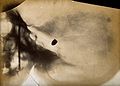
 A bullet in the base of a brain, viewed through x-ray. Photo Wellcome L0000624.jpg 1,190 × 1,614; 590 KB
-
 A bullet in the base of a brain, viewed through x-ray. Photo Wellcome V0029553.jpg 3,196 × 2,294; 2.58 MB
-
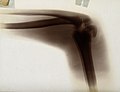
 A knee joint, viewed through x-ray. Photoprint from radiogra Wellcome V0029532.jpg 2,172 × 3,321; 3.06 MB
-
 A snake in the process of swallowing a small mammal, probabl Wellcome V0029551.jpg 2,300 × 3,172; 2.93 MB
-
 A snake in the process of swallowing a small mammal, probabl Wellcome V0029552.jpg 2,300 × 3,125; 2.36 MB
-
 Bullet in brain by Arthur Schuster.jpg 954 × 842; 268 KB
-
 Foot by Arthur Schuster.jpg 2,012 × 1,660; 1.14 MB
-
 Foot of Leonard Schuster.jpg 1,182 × 938; 381 KB
-
 Foot with needle by Arthur Schuster.jpg 1,504 × 1,158; 543 KB
-
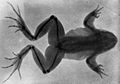 Frog by Arthur Schuster.jpg 1,394 × 978; 527 KB
-
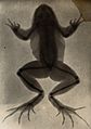 Frog by Arthur Schuster2.jpg 1,935 × 2,749; 1.81 MB
-
 Hand by Arthur Schuster.jpg 2,912 × 2,020; 2.1 MB
-
 Hand by Arthur Schuster2.jpg 1,900 × 2,628; 1.34 MB
-
 Hand with ring by Arthur Schuster.jpg 2,362 × 3,267; 2.76 MB
-
 Hands by Arthur Schuster.jpg 1,192 × 1,582; 739 KB
-
 Radiograph; foot of a pantomime artist. Wellcome L0000623EB.jpg 1,145 × 1,639; 523 KB
-
 Radiograph; foot of Leonard Schuster, aged six and a half. Wellcome L0000621EB.jpg 1,570 × 1,190; 579 KB
-
 Radiograph; frog with a broken leg that has healed. Wellcome L0000620EA.jpg 1,626 × 1,182; 708 KB
-
 Solar eclipse 1875Apr06 Lockyer.png 486 × 703; 258 KB
-
 The bones in the joint of an elbow, viewed through x-ray. Ph Wellcome V0029535.jpg 2,376 × 3,198; 2.74 MB
-
 The bones of a foot, possibly with leprosy, viewed through x Wellcome V0029544.jpg 3,224 × 2,404; 2.99 MB
-
 The bones of a foot, viewed through x-ray. Photoprint from r Wellcome V0029529.jpg 3,240 × 2,392; 2.83 MB
-
 The bones of a foot, viewed through x-ray. Photoprint from r Wellcome V0029537.jpg 2,340 × 3,132; 2.23 MB
-
 The bones of a forearm, viewed through x-ray. Photoprint fro Wellcome V0029545.jpg 2,396 × 3,276; 2.43 MB
-
 The bones of a frog, viewed through x-ray; revealing a heali Wellcome V0029531.jpg 2,320 × 3,232; 2.84 MB
-
 The bones of a hand and wrist, viewed through x-ray. Photopr Wellcome V0029543.jpg 3,150 × 2,285; 2.65 MB
-
 The bones of a hand, viewed through x-ray. Photoprint from r Wellcome V0029530.jpg 2,400 × 2,996; 2.32 MB
-
 The bones of a hand, viewed through x-ray. Photoprint from r Wellcome V0029536.jpg 2,446 × 3,162; 3.4 MB
-
 The bones of a hand, viewed through x-ray. Photoprint from r Wellcome V0029550.jpg 2,350 × 3,084; 3.36 MB
-
 The bones of a hand, viewed through x-ray; possibly with a n Wellcome V0029533.jpg 3,125 × 2,244; 2.8 MB
-
 The bones of a hand, with a ring on one finger, viewed throu Wellcome V0029534.jpg 2,362 × 3,282; 3.12 MB
-
 The bones of a hand, with the tip of the index finger missin Wellcome V0029542.jpg 2,340 × 3,126; 2.53 MB
-
 The bones of a knee joint of a young boy, viewed through x-r Wellcome V0029546.jpg 2,400 × 3,186; 2.25 MB
-
 The bones of a pantomime artist's foot, viewed through x-ray Wellcome L0014161.jpg 1,618 × 1,212; 485 KB
-
 The bones of a pantomime artist's foot, viewed through x-ray Wellcome L0016291.jpg 1,564 × 1,208; 516 KB
-
 The bones of a pantomime artist's foot, viewed through x-ray Wellcome V0029527.jpg 2,270 × 3,102; 2.09 MB
-
 The bones of a pantomime artist's foot, viewed through x-ray Wellcome V0029528.jpg 3,350 × 2,106; 2.12 MB
-
 The bones of a shoulder joint and ribs, viewed through x-ray Wellcome V0029547.jpg 2,416 × 3,232; 3.39 MB
-
 The bones of an elbow joint, viewed through x-ray. Photoprin Wellcome V0029549.jpg 2,388 × 3,220; 3.23 MB
-
 The bones of the ribs and vertebral column, viewed through x Wellcome V0029538.jpg 2,334 × 3,228; 2.57 MB
-
 The bones of the ribs and vertebral column, viewed through x Wellcome V0029548.jpg 2,334 × 3,162; 3.18 MB
-
 The bones of the wrist, viewed through x-ray. Photoprint fro Wellcome V0029539.jpg 2,340 × 3,160; 2.38 MB
-
 The fracture and dislocation of bones in an elbow joint, vie Wellcome V0029541.jpg 3,024 × 2,308; 2.09 MB
-
 Two hands, viewed through x-ray. Photoprint from radiograph Wellcome L0013210.jpg 1,298 × 1,590; 759 KB
-

-
 Two hands, viewed through x-ray. Photoprint from radiograph Wellcome V0029554.jpg 2,400 × 2,933; 2.08 MB


.jpg)
