Category:Image analysis
Jump to navigation
Jump to search
extraction of information from images via digital image processing techniques | |||||
| Upload media | |||||
| Subclass of | |||||
|---|---|---|---|---|---|
| Different from | |||||
| |||||
Subcategories
This category has the following 4 subcategories, out of 4 total.
Media in category "Image analysis"
The following 80 files are in this category, out of 80 total.
-
-
-
-
-
-
A-versatile-pipeline-for-the-multi-scale-digital-reconstruction-and-quantitative-analysis-of-3D-elife-11214-media2.ogv 1 min 24 s, 868 × 720; 28.42 MB
-
-
-
-
-
-
-
-
-
-
-
Basic flow through diag on white.png 649 × 294; 19 KB
-
BioSig3D-High-Content-Screening-of-Three-Dimensional-Cell-Culture-Models-pone.0148379.s005.ogv 1 min 2 s, 976 × 720; 4.96 MB
-
BioSig3D-High-Content-Screening-of-Three-Dimensional-Cell-Culture-Models-pone.0148379.s006.ogv 3 min 39 s, 976 × 720; 13.5 MB
-
BioSig3D-High-Content-Screening-of-Three-Dimensional-Cell-Culture-Models-pone.0148379.s007.ogv 1 min 37 s, 964 × 720; 6 MB
-
BioSig3D-High-Content-Screening-of-Three-Dimensional-Cell-Culture-Models-pone.0148379.s008.ogv 2 min 12 s, 964 × 720; 10.87 MB
-
BioSig3D-High-Content-Screening-of-Three-Dimensional-Cell-Culture-Models-pone.0148379.s009.ogv 2 min 19 s, 964 × 720; 11.7 MB
-
BioSig3D-High-Content-Screening-of-Three-Dimensional-Cell-Culture-Models-pone.0148379.s010.ogv 3 min 19 s, 1,106 × 720; 21.04 MB
-
BioSig3D-High-Content-Screening-of-Three-Dimensional-Cell-Culture-Models-pone.0148379.s011.ogv 6.0 s, 318 × 333; 58 KB
-
BioSig3D-High-Content-Screening-of-Three-Dimensional-Cell-Culture-Models-pone.0148379.s012.ogv 6.2 s, 293 × 333; 51 KB
-
BioSig3D-High-Content-Screening-of-Three-Dimensional-Cell-Culture-Models-pone.0148379.s013.ogv 8.6 s, 320 × 333; 199 KB
-
BioSig3D-High-Content-Screening-of-Three-Dimensional-Cell-Culture-Models-pone.0148379.s014.ogv 8.0 s, 382 × 333; 354 KB
-
BisQue user interface.png 1,316 × 902; 691 KB
-
Bumblebee-Homing-The-Fine-Structure-of-Head-Turning-Movements-pone.0135020.s001.ogv 1 min 10 s, 1,280 × 720; 9.96 MB
-
Cardamom buns image vector scope plot color.svg 900 × 900; 814 KB
-
Delta Calculation.png 486 × 341; 20 KB
-
-
Empresas del Grupo RPP.jpg 650 × 714; 80 KB
-
Enhanced-Vitreous-Imaging-in-Healthy-Eyes-Using-Swept-Source-Optical-Coherence-Tomography-pone.0102950.s001.ogv 22 s, 1,500 × 680; 34.09 MB
-
Enhanced-Vitreous-Imaging-in-Healthy-Eyes-Using-Swept-Source-Optical-Coherence-Tomography-pone.0102950.s002.ogv 24 s, 1,500 × 500; 27.27 MB
-
-
-
Enhanced-Vitreous-Imaging-in-Healthy-Eyes-Using-Swept-Source-Optical-Coherence-Tomography-pone.0102950.s005.ogv 22 s, 1,500 × 700; 47.24 MB
-
Enhanced-Vitreous-Imaging-in-Healthy-Eyes-Using-Swept-Source-Optical-Coherence-Tomography-pone.0102950.s006.ogv 14 s, 1,500 × 500; 29.51 MB
-
Example of flow in a region.png 675 × 360; 160 KB
-
Example of object tracking.png 739 × 278; 135 KB
-
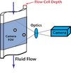
Flow cell Cross Section.png 812 × 961; 33 KB
-
HCS overview cs.png 3,794 × 1,967; 5.71 MB
-
HCS overview.png 3,794 × 1,967; 5.7 MB
-
High-Content-Screening-in-Zebrafish-Embryos-Identifies-Butafenacil-as-a-Potent-Inducer-of-Anemia-pone.0104190.s034.ogv 3.0 s, 1,392 × 1,040; 2.01 MB
-
High-Content-Screening-in-Zebrafish-Embryos-Identifies-Butafenacil-as-a-Potent-Inducer-of-Anemia-pone.0104190.s035.ogv 3.0 s, 1,392 × 1,040; 2.23 MB
-
High-Speed-Imaging-of-Cavitation-around-Dental-Ultrasonic-Scaler-Tips-pone.0149804.s001.ogv 6.1 s, 543 × 512; 3.76 MB
-
High-Speed-Imaging-of-Cavitation-around-Dental-Ultrasonic-Scaler-Tips-pone.0149804.s002.ogv 30 s, 128 × 80; 450 KB
-
High-Speed-Imaging-of-Cavitation-around-Dental-Ultrasonic-Scaler-Tips-pone.0149804.s003.ogv 10 s, 384 × 128; 1.1 MB
-
High-Speed-Imaging-of-Cavitation-around-Dental-Ultrasonic-Scaler-Tips-pone.0149804.s004.ogv 10 s, 384 × 128; 1.41 MB
-
High-Speed-Imaging-of-Cavitation-around-Dental-Ultrasonic-Scaler-Tips-pone.0149804.s005.ogv 10 s, 384 × 128; 1.26 MB
-
Image analysis.png 1,192 × 541; 672 KB
-
Image-analysis-flow.png 781 × 731; 220 KB
-
Inhomogeneity-Based-Characterization-of-Distribution-Patterns-on-the-Plasma-Membrane-pcbi.1005095.s017.ogv 5.0 s, 1,450 × 1,743; 1.16 MB
-
Interpretation of aerial photographs (IA interpretationof00cris).pdf 1,206 × 1,612, 56 pages; 2.34 MB
-
-
-
-
-
-
-
-
-
-
Loch Ness Rines 1975.png 758 × 575; 241 KB
-
Mansi Plesiosaur 1977.png 758 × 567; 446 KB
-
Object based image analysis.jpg 1,500 × 1,200; 2.53 MB
-
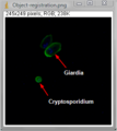
Object-registration-example.png 272 × 305; 18 KB
-
Parallactic Instrument of Kapteyn and Glass Plate Holder.jpg 1,328 × 930; 72 KB
-
Parallactic Instrument of Kapteyn.tif 2,848 × 4,288; 34.95 MB
-
ParallacticInstrumentKapteynDrawing.png 1,063 × 709; 117 KB
-
Phoretix 1D - Automatic Band Detection.png 2,560 × 1,371; 933 KB
-
Semantic Gap.svg 990 × 315; 41 KB
-
Shearing operation.png 509 × 198; 63 KB
-
Spinning dancer explained.png 600 × 400; 58 KB
-
Visual-parameter-optimisation-for-biomedical-image-processing-1471-2105-16-S11-S9-S1.ogv 3 min 3 s, 960 × 540; 9.49 MB
-
-


