Category:Human tibia
Jump to navigation
Jump to search
Subcategories
This category has the following 16 subcategories, out of 16 total.
- SVG human tibia (4 F)
3
- 3D data of human tibia (2 F)
A
D
- Daniel Sickles's leg (7 F)
F
M
O
P
S
- Low kicking (10 F)
- Shin guards (68 F)
V
- Videos of human tibia (3 F)
X
Media in category "Human tibia"
The following 143 files are in this category, out of 143 total.
-
3D CT Reconstruction of Distal tibia fracture.gif 988 × 918; 9.32 MB
-
American quarterly of roentgenology (1912) (14570606359).jpg 2,144 × 2,894; 2.09 MB
-
BodyParts3D Tibia.stl 5,120 × 2,880; 165 KB
-
BrodieAbscessRadiograph.jpg 644 × 1,654; 70 KB
-
Cro-Magnon-Modern Human Tibia comarision.png 777 × 408; 49 KB
-
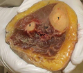
Cross section of cadaver limb.png 1,200 × 1,047; 1.61 MB
-
Cunningham’s Text-book of Anatomy (1914) - Fig 248.png 1,250 × 913; 1.01 MB
-
Cunningham’s Text-book of Anatomy (1914) - Fig 249.png 1,164 × 1,182; 1.07 MB
-
Cunningham’s Text-book of Anatomy (1914) - Fig 251.png 752 × 2,240; 1,019 KB
-
Cunningham’s Text-book of Anatomy (1914) - Fig 363.png 1,014 × 1,545; 1.42 MB
-
Cunningham’s Text-book of Anatomy (1914) - Fig 384.png 572 × 2,563; 1.11 MB
-
De-Tibiakopf.ogg 2.2 s; 21 KB
-
Dixon's Manual of human osteology (1912) - Fig 071.png 846 × 2,235; 754 KB
-
Dixon's Manual of human osteology (1912) - Fig 072.png 912 × 2,205; 689 KB
-
Dixon's Manual of human osteology (1912) - Fig 073.png 1,426 × 872; 859 KB
-
Dixon's Manual of human osteology (1912) - Fig 074.png 852 × 1,869; 642 KB
-
Dixon's Manual of human osteology (1912) - Fig 075.png 909 × 2,019; 673 KB
-
Fersensporn.jpg 1,112 × 692; 41 KB
-
Fibula et tibia.png 475 × 762; 95 KB
-
Fracture of a tibia, five weeks after the accident (Fig 3) Wellcome L0062605.jpg 5,024 × 4,152; 4.45 MB
-
Fracture of a tibia, five weeks after the accident (Figs 1-2) Wellcome L0062604.jpg 5,556 × 4,400; 4.49 MB
-
Gerrish's Text-book of Anatomy (1902) - Fig. 132.png 459 × 1,953; 543 KB
-
Gerrish's Text-book of Anatomy (1902) - Fig. 190.png 862 × 578; 527 KB
-
Gerrish's Text-book of Anatomy (1902) - Fig. 191.png 972 × 2,037; 810 KB
-
Gerrish's Text-book of Anatomy (1902) - Fig. 192.png 908 × 2,000; 597 KB
-
Gerrish's Text-book of Anatomy (1902) - Fig. 193.png 957 × 2,211; 836 KB
-
Gerrish's Text-book of Anatomy (1902) - Fig. 194.png 872 × 2,016; 766 KB
-
Gerrish's Text-book of Anatomy (1902) - Fig. 195.png 1,128 × 636; 224 KB
-
Gray257.png 324 × 324; 27 KB
-
Gray258.png 346 × 1,000; 46 KB
-
Gray259 he.png 377 × 1,000; 39 KB
-
Gray259.png 367 × 1,000; 43 KB
-
Gray260.png 344 × 450; 13 KB
-
Gray261.png 231 × 400; 15 KB
-
Gray354.png 550 × 424; 39 KB
-
Gray355.png 600 × 487; 57 KB
-
Gray356.png 434 × 400; 30 KB
-
Gray357-ar.png 550 × 469; 153 KB
-
Gray357.png 550 × 469; 46 KB
-
Gray440 color.png 600 × 413; 107 KB
-
Gray440.png 600 × 413; 144 KB
-
Gunshot injury to the tibia.jpg 438 × 1,081; 188 KB
-
Holden's human osteology (1899) - Plt35 Fig01-02.png 993 × 1,719; 874 KB
-
Holden's human osteology (1899) - Plt35 Fig03.png 870 × 1,749; 644 KB
-
Human tibia.stl 5,120 × 2,880; 7.07 MB
-
J.F. Gautier D'Agoty, Myologie complette en coleur... Wellcome L0023743.jpg 1,186 × 1,538; 636 KB
-
Jud-sune.gif 430 × 430; 5 KB
-
K-Knie-z2.jpg 2,083 × 1,960; 236 KB
-
Left tibia - close up - animation.gif 450 × 450; 553 KB
-
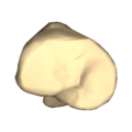
Left tibia - close up - anterior view.png 4,500 × 4,500; 532 KB
-
Left tibia - close up - inferior view.png 4,500 × 4,500; 1.64 MB
-
Left tibia - close up - lateral view.png 4,500 × 4,500; 539 KB
-
Left tibia - close up - medial view.png 4,500 × 4,500; 554 KB
-
Left tibia - close up - posterior view.png 4,500 × 4,500; 557 KB
-
Left tibia - close up - superior view.png 4,500 × 4,500; 1.36 MB
-
Merkel's Human Anatomy (1913) - Vol 3 - Fig 123-124.png 1,295 × 2,061; 509 KB
-
Merkel's Human Anatomy (1913) - Vol 3 - Fig 125-126.png 1,323 × 1,932; 424 KB
-
Morris' human anatomy (1898) - Fig 163.png 1,616 × 2,528; 1.7 MB
-
Morris' human anatomy (1898) - Fig 164.png 1,596 × 2,576; 1.46 MB
-
Morris' human anatomy (1933) - Fig 273.png 2,007 × 3,046; 2.46 MB
-
Morris' human anatomy (1933) - Fig 274.png 1,990 × 3,006; 2.29 MB
-
Necrosis of the tibia with sequestrum Wellcome L0061260.jpg 6,256 × 2,388; 2.15 MB
-
Osteomyelitis of Tibia in Child.jpg 1,200 × 1,600; 606 KB
-
Ostermyelitis Tibia.jpg 1,200 × 1,600; 258 KB
-
Quain's elements of anatomy (1891) - Vol2 Part1- Fig 141.png 867 × 802; 888 KB
-
Quain's elements of anatomy (1891) - Vol2 Part1- Fig 216.png 1,083 × 563; 589 KB
-
Right tibia - close up - animation.gif 450 × 450; 554 KB
-
Right tibia - close up - anterior view.png 4,500 × 4,500; 515 KB
-
Right tibia - close up - inferior view.png 4,500 × 4,500; 1.53 MB
-
Right tibia - close up - lateral view.png 4,500 × 4,500; 555 KB
-
Right tibia - close up - medial view.png 4,500 × 4,500; 537 KB
-
Right tibia - close up - posterior view.png 4,500 × 4,500; 543 KB
-
Right tibia - close up - superior view.png 4,500 × 4,500; 1.35 MB
-
Scheletul membrului inferior 2.tif 180 × 517; 364 KB
-
Slide16C.JPG 960 × 720; 57 KB
-
Slide17C.JPG 960 × 720; 56 KB
-
Slide1Bebe.JPG 960 × 720; 71 KB
-
Slide1besa.JPG 960 × 720; 73 KB
-
Slide1DDDDD.JPG 960 × 720; 64 KB
-
Slide1dede.JPG 960 × 720; 53 KB
-
Slide1wewe.JPG 960 × 720; 77 KB
-
Slide2besa.JPG 960 × 720; 79 KB
-
Slide2cdcd-ar.jpg 960 × 720; 88 KB
-
Slide2cdcd.JPG 960 × 720; 55 KB
-
Slide2dede.JPG 960 × 720; 54 KB
-
Slide2wewew.JPG 960 × 720; 73 KB
-
Slide3dada.JPG 960 × 720; 64 KB
-
Slide3ecce.JPG 960 × 720; 71 KB
-
Slide4wewe.JPG 960 × 720; 63 KB
-
Slide8CEC7.JPG 960 × 720; 74 KB
-
Slide8DDDD.JPG 960 × 720; 58 KB
-
Sobo 1909 151.png 1,352 × 844; 3.27 MB
-
Sobo 1909 152.png 1,220 × 640; 2.24 MB
-
Sobo 1909 170.png 1,036 × 1,640; 4.87 MB
-
Tape20.png 379 × 487; 116 KB
-
Tape21.png 379 × 487; 250 KB
-
Tape22.png 758 × 487; 404 KB
-
Tape23.png 758 × 487; 416 KB
-
Testut's Treatise on Human Anatomy (1911) - Vol 1 - Fig 362-363-364.png 2,425 × 2,742; 1.88 MB
-
The Boxgrove Tibia.jpg 589 × 315; 26 KB
-
Tibia - animation left leg.gif 450 × 450; 528 KB
-
Tibia - animation right leg.gif 450 × 450; 530 KB
-
Tibia - animation.gif 450 × 450; 1.69 MB
-
Tibia - animation2.gif 450 × 450; 981 KB
-
Tibia - condylus lateralis et medialis.jpg 4,608 × 3,456; 4.8 MB
-
Tibia - detail of bone tissue (proximal end) 2.jpg 3,976 × 3,008; 4.44 MB
-
Tibia - detail of bone tissue (proximal end).jpg 4,608 × 3,456; 5.3 MB
-
Tibia - detail of malleolus medialis bone tissue.jpg 2,976 × 2,784; 2.98 MB
-
Tibia - dex, sin.jpg 4,608 × 3,456; 4.62 MB
-
Tibia - facies articularis superior (dex, sin).jpg 4,320 × 2,408; 3.4 MB
-
Tibia - facies articularis superior 2.jpg 2,932 × 2,108; 2.43 MB
-
Tibia - facies articularis superior.jpg 3,408 × 2,676; 3.42 MB
-
Tibia - frontal view.png 4,500 × 4,500; 2.78 MB
-
Tibia - frontal view2.png 4,500 × 4,500; 852 KB
-
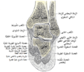
Tibia - inferior epiphysis (anterior view) Arabic YM.jpg 899 × 628; 161 KB
-
Tibia - inferior epiphysis (anterior view).jpg 960 × 720; 71 KB
-
Tibia - inferior epiphysis (posterior view).jpg 960 × 720; 54 KB
-
Tibia - lateral view.png 4,500 × 4,500; 1.63 MB
-
Tibia - lateral view2.png 4,500 × 4,500; 493 KB
-
Tibia - malleolus medialis (dex, sin).jpg 3,832 × 2,472; 3.2 MB
-
Tibia - malleolus medialis.jpg 4,608 × 3,456; 4.38 MB
-
Tibia - medial view - right leg.png 4,500 × 4,500; 478 KB
-
Tibia - posterior view.png 4,500 × 4,500; 2.69 MB
-
Tibia - posterior view2.png 4,500 × 4,500; 840 KB
-
Tibia - superior epiphysis (anterior view).jpg 960 × 720; 48 KB
-
Tibia - superior epiphysis (posterior view).jpg 960 × 720; 56 KB
-
Tibia - superior epiphysis (superior view).jpg 960 × 720; 87 KB
-
Tibia Anatomy by Jason Christian.webm 51 s, 1,280 × 720; 9.24 MB
-
Tibia and fibula bones. Ink and watercolour, 1830-1835?, aft Wellcome V0008213ER.jpg 1,179 × 1,714; 1.03 MB
-
Tibia and Fibula Overview.webm 3 min 14 s, 1,077 × 606; 12.69 MB
-
Tibia and Fibula Walk-thru by Bob Myers.webm 5 min 57 s, 640 × 480; 12.78 MB
-
Tibia bones; five figures. Pencil drawing, ca. 1809. Wellcome V0008238ER.jpg 1,396 × 2,248; 1.64 MB
-
Tibia bones; four figures. Pencil drawing, ca. 1804. Wellcome V0008237.jpg 2,484 × 3,096; 3.19 MB
-
Tibia bones; three figures. Pencil drawing, ca. 1809. Wellcome V0008238EL.jpg 1,413 × 2,238; 1.68 MB
-
Tibia derecha con lesión traumática.png 2,604 × 4,864; 3.13 MB
-
Tibia, fibula (anterior).jpg 3,860 × 1,696; 2.28 MB
-
Tibia-CT-Biomechanics-part2.png 1,119 × 701; 392 KB
-
Tibia-Plafond O - CR - 001.jpg 959 × 1,069; 143 KB
-
Tibia.jpg 4,384 × 2,648; 3.51 MB
-
Tibiae with treponematosis.png 850 × 1,212; 1.57 MB
-
Tibiaumstellung bei Paget.jpg 926 × 1,310; 419 KB


