Category:Human pituitary gland
Jump to navigation
Jump to search
Subcategories
This category has the following 11 subcategories, out of 11 total.
Media in category "Human pituitary gland"
The following 94 files are in this category, out of 94 total.
-
01S-3A 022 ActivBil.ContraceptionHormonale.SchFoncMuet2.svg 1,286 × 909; 80 KB
-
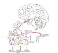
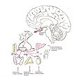
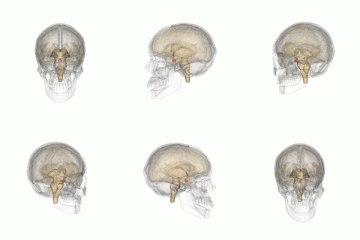
201405 pituitary gland in brain.png 400 × 400; 31 KB
-
201405 pituitary gland.png 400 × 400; 36 KB
-
A-GRFa2Prop1Stem-(GPS)-Cell-Niche-in-the-Pituitary-pone.0004815.s005.ogv 15 s, 768 × 576; 1.72 MB
-
A-GRFa2Prop1Stem-(GPS)-Cell-Niche-in-the-Pituitary-pone.0004815.s006.ogv 15 s, 768 × 576; 3.35 MB
-
A-GRFa2Prop1Stem-(GPS)-Cell-Niche-in-the-Pituitary-pone.0004815.s007.ogv 17 s, 768 × 576; 1.09 MB
-
A-GRFa2Prop1Stem-(GPS)-Cell-Niche-in-the-Pituitary-pone.0004815.s008.ogv 1 min 22 s, 768 × 576; 4.09 MB
-
A-GRFa2Prop1Stem-(GPS)-Cell-Niche-in-the-Pituitary-pone.0004815.s009.ogv 1 min 5 s, 768 × 576; 2.91 MB
-
A-GRFa2Prop1Stem-(GPS)-Cell-Niche-in-the-Pituitary-pone.0004815.s010.ogv 33 s, 768 × 576; 2.39 MB
-
A-GRFa2Prop1Stem-(GPS)-Cell-Niche-in-the-Pituitary-pone.0004815.s011.ogv 20 s, 700 × 524; 2.56 MB
-
Anatomy of the cavernous sinus.jpg 800 × 499; 93 KB
-
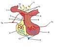
Anterior and posterior pituitary.jpg 630 × 240; 28 KB
-
Aufgabe der Hypophyse.svg 512 × 542; 187 KB
-
Basel 2012-10-05 Batch 2 (26).JPG 2,736 × 3,648; 3.03 MB
-
Basel 2012-10-05 Batch 2 (27).JPG 2,736 × 3,648; 2.58 MB
-
Basel 2012-10-05 Batch 2 (28).JPG 3,648 × 2,736; 3.68 MB
-
Biosintesi della melatonina.JPG 378 × 960; 32 KB
-
Corticotroph traces.png 2,481 × 1,057; 157 KB
-
De-Hypophyse.ogg 2.2 s; 21 KB
-
Development of the pituitary gland.webm 1 min 26 s, 640 × 480; 1.42 MB
-
Emergent-Synchronous-Bursting-of-Oxytocin-Neuronal-Network-pcbi.1000123.s001.ogv 20 s, 560 × 420; 1.85 MB
-
Endocrine growth regulation.png 1,698 × 2,080; 1.05 MB
-
Figure 28 01 07.JPG 1,058 × 713; 250 KB
-
Gray1180-ar.png 672 × 375; 165 KB
-
Gray1180.png 672 × 375; 45 KB
-
Gray1181-ar.png 575 × 350; 127 KB
-
Gray1181.png 575 × 350; 34 KB
-
Gray516.png 600 × 681; 113 KB
-
Gray721.png 600 × 333; 59 KB
-
Gray994 zh.png 600 × 861; 329 KB
-
Gray994.png 600 × 861; 112 KB
-
Grays pituitary.png 600 × 333; 65 KB
-
Hipofisis-hormonas.jpg 896 × 814; 77 KB
-
Hipófise - Posterior.png 555 × 401; 174 KB
-
Hipófise ou Glândula Pituitária.jpg 289 × 174; 7 KB
-
Hipófisis-hormonas2.jpg 896 × 814; 80 KB
-
Hormonas-hipofisis.jpg 896 × 814; 77 KB
-
Hormone in der Pubertät.svg 512 × 390; 217 KB
-
Human brain dura mater (reflections) description.JPG 700 × 486; 49 KB
-
Human brain left midsagitttal view closeup description 2.JPG 701 × 490; 61 KB
-
Hypofýza a pineální žláza.svg 114 × 76; 59 KB
-
Hypophyse 2M - MR - 001.jpg 1,467 × 1,881; 292 KB
-
Hypophyse 31jw postpartum - MRT T1 sag - 001.jpg 1,535 × 1,318; 123 KB
-
Hypophyse et glandes pinéales.gif 400 × 259; 19 KB
-
Hypophyse MRT sag.png 1,198 × 1,180; 615 KB
-
Hypophyse und Epiphyse.jpg 656 × 512; 74 KB
-
Hypophyse.png 2,252 × 2,072; 692 KB
-
Hypophyseal gland.jpg 960 × 720; 87 KB
-
Hypophysis.jpg 467 × 304; 16 KB
-
Hypophysis3.gif 575 × 350; 39 KB
-
Illu pituitary pineal glands ja.JPG 400 × 255; 23 KB
-
Illu pituitary pineal glands zh.jpg 400 × 259; 31 KB
-
Illu pituitary pineal glands-az.png 400 × 259; 84 KB
-
Illu pituitary pineal glands.jpg 400 × 259; 23 KB
-
Innvervazione dell'epifisi.JPG 567 × 567; 27 KB
-
Interplay between the central and peripheral circadian clocks.jpg 766 × 1,221; 127 KB
-
L03 infundibulum 001.jpg 378 × 377; 30 KB
-
Location of hypothalamus, pituitary gland and olfactory bulb. .gif 440 × 243; 14 KB
-
LocationOfHypothalamus.jpg 350 × 250; 21 KB
-
Nasennebenhöhlen.gif 600 × 428; 66 KB
-
Neurohipófisis.jpg 832 × 1,151; 166 KB
-
Pineal Gland and Pituitary Body.jpg 291 × 170; 10 KB
-
Pituitary development animation.gif 300 × 200; 52 KB
-
Pituitary gland et vessel.jpg 700 × 600; 57 KB
-
Pituitary gland image.png 800 × 455; 308 KB
-
Pituitary gland representation.PNG 261 × 236; 8 KB
-
Pituitary gland small.gif 200 × 200; 592 KB
-
Pituitary gland-optic chiasm-sella turcica-es.png 625 × 347; 69 KB
-
Pituitary gland-optic chiasm-sella turcica.jpg 600 × 333; 127 KB
-
Pituitary gland.png 468 × 332; 210 KB
-
Pituitary pineal glands.jpg 355 × 239; 22 KB
-
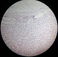
Pituitary slide.jpg 2,993 × 2,908; 3.51 MB
-
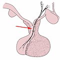
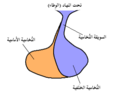
Pituitary Stalk-ar.png 309 × 257; 9 KB
-
Pituitary Stalk.png 309 × 257; 8 KB
-
Pituitary Tumor Removal.png 1,500 × 750; 877 KB
-
Pituitary xanthogranuloma.jpg 2,080 × 1,542; 857 KB
-
Prolactinoma-art-zh.jpg 261 × 358; 50 KB
-
Prolactinoma-art.jpg 261 × 358; 70 KB
-
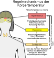
Regelmechanismus Körpertemperatur.svg 512 × 542; 240 KB
-
Sajous's analytical cyclopædia of practical medicine (1904) (14778360432).jpg 1,554 × 2,282; 574 KB
-
Sella mri-contra.jpg 1,100 × 600; 112 KB
-
Sheehans Syndrome.webm 5 min 10 s, 1,920 × 1,080; 34.16 MB
-
Silla turca TalloHipof.png 512 × 322; 309 KB
-
Sistema porta H-H.jpg 1,226 × 716; 170 KB
-
Sleep.PNG 537 × 455; 35 KB
-
Slide2STE.JPG 960 × 720; 106 KB
-
Sobo 1909 624 ar.png 3,060 × 2,247; 5.38 MB
-
Sobo 1909 624.png 3,060 × 2,247; 19.71 MB
-
ThyroidImage.jpg 400 × 320; 68 KB
-
Voluminoese Hypophyse in der Schwangerschaft 30W - MR T1 - 001.jpg 3,633 × 1,380; 280 KB
-
Схема гипоталамо-эпифизарной системы.jpg 719 × 720; 145 KB


