Category:Human olfactory bulb
Jump to navigation
Jump to search
Media in category "Human olfactory bulb"
The following 57 files are in this category, out of 57 total.
-
1543, Andreas Vesalius' Fabrica, Base Of The Brain.jpg 1,202 × 1,346; 1.11 MB
-
1543,Vesalius'OlfactoryBulbs.jpg 290 × 308; 24 KB
-
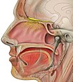
Anatomy of the human nasal cavity.png 642 × 665; 329 KB
-
Anatomy, physiology and hygiene (1900) (14756274196).jpg 1,468 × 1,140; 657 KB
-
Autopsy brain.jpg 1,600 × 1,059; 328 KB
-
Blausen 0111 BrainLobes.png 2,250 × 1,350; 1.55 MB
-
Blausen 0284 CranialNerves esp.jpg 2,250 × 1,350; 902 KB
-
Blausen 0284 CranialNerves.png 2,250 × 1,350; 1.82 MB
-
Brain human normal inferior view with labels ar.png 424 × 505; 362 KB
-
Brain human normal inferior view with labels ar.svg 424 × 505; 244 KB
-
Brain human normal inferior view with labels en.svg 424 × 505; 172 KB
-
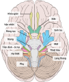
Brain human normal inferior view with labels vi.svg 424 × 505; 262 KB
-
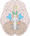
Brain human normal inferior view.svg 424 × 505; 204 KB
-
Cerebral Gyri - Inferior Surface2.png 757 × 569; 431 KB
-
Cribriform plate and Olfactory nerve - animation.gif 600 × 600; 8.08 MB
-
Cribriform plate and Olfactory nerve - superior view.svg 1,500 × 1,500; 904 KB
-
Early Olfactory System.svg 1,017 × 1,469; 81 KB
-
Figure 07 - Mitral cell development (Flat).png 9,787 × 5,673; 3.42 MB
-
Fnsys-05-00062-g003.jpg 491 × 165; 80 KB
-
Gray724.png 600 × 588; 73 KB
-
Gray755.png 500 × 329; 46 KB
-
Gray772 vi.png 400 × 328; 37 KB
-
Gray772.png 400 × 328; 13 KB
-
Gray780.png 650 × 480; 68 KB
-
Gray858.png 450 × 422; 43 KB
-
Head olfactory nerve - olfactory bulb en.png 743 × 824; 835 KB
-
Head olfactory nerve - olfactory bulb ja.jpg 725 × 825; 515 KB
-
Head Olfactory Nerve Labeled bn.png 1,662 × 1,472; 2.66 MB
-
Head Olfactory Nerve Labeled vi.png 957 × 879; 984 KB
-
Head Olfactory Nerve Labeled-es.png 957 × 883; 1.15 MB
-
Head Olfactory Nerve Labeled.png 957 × 879; 928 KB
-
Head olfactory nerve.jpg 725 × 825; 529 KB
-
Human brain anterior-inferior view description.JPG 330 × 475; 31 KB
-
Human brain anterior-inferior view.JPG 330 × 475; 28 KB
-
Human brain inferior view description 2.JPG 373 × 467; 36 KB
-
Human brain inferior view.JPG 373 × 467; 34 KB
-
Human brainstem anterior view 2 description.JPG 346 × 487; 35 KB
-
Location of hypothalamus, pituitary gland and olfactory bulb. .gif 440 × 243; 14 KB
-
Location of olfactory ensheathing cells (OECs) within the olfactory system.png 1,860 × 1,357; 860 KB
-
-
Olfactory system.svg 1,151 × 620; 434 KB
-
Posterior Superior Nasal Nerve.png 724 × 697; 418 KB
-
-
Slide1ee.JPG 960 × 720; 137 KB
-
Slide21ior.JPG 960 × 720; 115 KB
-
Slide2Dsa.JPG 960 × 720; 89 KB
-
Slide2MIR.JPG 960 × 720; 115 KB
-
Slide2PIT.JPG 960 × 720; 97 KB
-
Slide2STE.JPG 960 × 720; 106 KB
-
Sobo 1909 623 ar.png 2,404 × 2,652; 5.62 MB
-
Sobo 1909 623.png 2,404 × 2,652; 18.27 MB
-
Sobo 1909 634.png 1,089 × 703; 2.19 MB
-
Sobo 1909 647.png 1,168 × 1,165; 3.9 MB
-
Structures of brain.jpg 935 × 648; 104 KB
-
TE-Nose diagram.svg 800 × 738; 284 KB
-
Training.seer.cancer.gov - illu cranial nerves1.jpg 381 × 442; 35 KB
-
ヒトの嗅覚器の構造.svg 800 × 800; 26 KB


