Category:Human metacarpus
Jump to navigation
Jump to search
| Human hand bones |

 Human anatomy Metacarpal bones
| ||
|---|---|---|
Subcategories
This category has the following 8 subcategories, out of 8 total.
1
2
- Second metacarpal bone (21 F)
3
- Third metacarpal bone (23 F)
4
- Fourth metacarpal bone (19 F)
5
- Fifth metacarpal bone (27 F)
C
F
H
Media in category "Human metacarpus"
The following 94 files are in this category, out of 94 total.
-
 American quarterly of roentgenology (1909) (14570807748).jpg 2,512 × 4,044; 604 KB
-
 Bidloo Ontleding 1690 71.jpg 1,200 × 1,614; 322 KB
-
 Braus 1921 187.png 1,622 × 1,544; 7.18 MB
-
 Braus 1921 201.png 1,660 × 816; 1.08 MB
-
 Braus 1921 216.png 1,575 × 822; 3.71 MB
-
 Carpal-boss-3d.jpg 918 × 1,095; 179 KB
-
 Carpus.jpg 960 × 720; 80 KB
-
 Carpus2.jpg 960 × 720; 79 KB
-
 Cunningham’s Text-book of Anatomy (1914) - Fig 210.png 1,434 × 1,071; 1,009 KB
-
 Cunningham’s Text-book of Anatomy (1914) - Fig 211.png 1,398 × 1,068; 900 KB
-
 Cunningham’s Text-book of Anatomy (1914) - Fig 221.png 608 × 868; 788 KB
-
 Cunningham’s Text-book of Anatomy (1914) - Fig 228.png 1,662 × 706; 825 KB
-
 Cunningham’s Text-book of Anatomy (1914) - Fig 351.png 1,863 × 1,586; 1.78 MB
-
 Cunningham’s Text-book of Anatomy (1914) - Fig 353.png 1,854 × 1,191; 773 KB
-
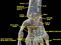
 Dissection of the human hand - 01.jpg 960 × 720; 81 KB
-
 Dissection of the human hand - 02.jpg 960 × 720; 88 KB
-
 Dixon's Manual of human osteology (1912) - Fig 048.png 1,452 × 1,248; 636 KB
-
 Dixon's Manual of human osteology (1912) - Fig 049.png 1,478 × 1,264; 749 KB
-
 Dixon's Manual of human osteology (1912) - Fig 050.png 1,155 × 1,146; 483 KB
-
 Dixon's Manual of human osteology (1912) - Fig 051.png 1,198 × 1,242; 737 KB
-
 Dixon's Manual of human osteology (1912) - Fig 052.png 1,116 × 1,815; 652 KB
-
 Dixon's Manual of human osteology (1912) - Fig 053.png 1,530 × 1,653; 964 KB
-
 Dixon's Manual of human osteology (1912) - Fig 054.png 1,392 × 1,605; 817 KB
-
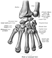
 Gray219 - Metacarpus.png 650 × 831; 285 KB
-
 Gray219.png 650 × 831; 76 KB
-
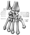
 Gray220 - Metacarpus.png 733 × 900; 272 KB
-
 Gray220.png 733 × 900; 73 KB
-
 Gray234.png 400 × 594; 32 KB
-
 Gray334.png 509 × 466; 49 KB
-
 Gray335.png 508 × 441; 46 KB
-
 Gray336.png 550 × 430; 48 KB
-
 Guanajuato mummy 02.jpg 1,000 × 800; 368 KB
-
 Haishu.png 1,380 × 780; 237 KB
-
 Hand-bones.jpg 1,324 × 1,107; 100 KB
-
 Handskelett 55j weiblich.png 562 × 1,084; 275 KB
-
 Handskelett 66j maennlich.png 638 × 1,088; 343 KB
-
 Handwurzel und Mittelhand 19j weiblich.png 716 × 626; 311 KB
-
 Medical X-Ray imaging MFM05 nevit.jpg 1,784 × 2,384; 1.57 MB
-
 Medical X-Ray imaging MYO06 nevit.jpg 1,784 × 2,384; 428 KB
-
 Medical X-Ray imaging OCU06 nevit.jpg 1,784 × 2,384; 463 KB
-
 Medical X-Ray imaging VKK07 nevit.jpg 1,784 × 2,384; 727 KB
-
 Medical X-Ray imaging VNH07 nevit.jpg 1,974 × 2,486; 704 KB
-
 Medical X-Ray imaging XGT07 nevit.jpg 2,486 × 1,974; 1.05 MB
-
 Metacarpal bones (left hand) - animation01.gif 450 × 450; 1.56 MB
-
 Metacarpal bones (left hand) - animation02.gif 450 × 450; 3.25 MB
-
 Metacarpal bones (left hand) 01 palmar view with label.png 4,500 × 4,500; 1.95 MB
-
 Metacarpal bones (left hand) 01 palmer view.png 4,500 × 4,500; 1.61 MB
-
 Metacarpal bones (left hand) 02 dorsal view.png 4,500 × 4,500; 1.49 MB
-
 Metacarpal bones (left hand) 03 ulnar view.png 4,500 × 4,500; 1.42 MB
-
 Metacarpal bones (left hand) 04 radial view.png 4,500 × 4,500; 1.36 MB
-
 Metacarpal bones (left hand) 05 palmar view.png 4,500 × 4,500; 1.37 MB
-
 Metacarpal bones (left hand) 06 dorsal view.png 4,500 × 4,500; 1.26 MB
-
 Metacarpal bones - animation01.gif 450 × 450; 2.59 MB
-
 Metacarpal bones 01 palmer view.png 4,500 × 4,500; 3.49 MB
-
 Metacarpal bones 02 dorsal view.png 4,500 × 4,500; 3.62 MB
-
 Metacarpal bones 03 radial view.png 4,500 × 4,500; 1.8 MB
-
 Metacarpal bones2.jpg 960 × 720; 60 KB
-
 Metacarpal bones3.jpg 960 × 720; 84 KB
-
 Metacarpals numbered-en (left hand).svg 699 × 544; 176 KB
-
 Metacarpals numbered-en.svg 699 × 544; 160 KB
-
 Metacarpus (left hand) dorsal view.png 960 × 720; 724 KB
-
 Metacarpus ant with label.png 1,024 × 597; 851 KB
-
 Metacarpus ant.jpg 1,024 × 597; 84 KB
-
 Metacarpus med.jpg 1,024 × 511; 77 KB
-
 NECK METACARPAL.png 416 × 596; 69 KB
-
 Ospoignet.gif 550 × 430; 56 KB
-
 Q11.jpg 960 × 720; 93 KB
-
 Radius-epiphysiolyse01.jpg 366 × 619; 41 KB
-
 RightHumanAnteriorDistalRadiusUlnaCarpals.jpg 2,661 × 1,681; 687 KB
-
 Scheme human hand bones-en.svg 406 × 391; 144 KB
-
 Scheme human hand bones-zh.svg 406 × 391; 113 KB
-
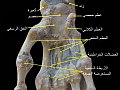
 Slide1dsds-ar.jpg 960 × 720; 139 KB
-
 Slide1dsds.JPG 960 × 720; 67 KB
-
 Slide1tyty.JPG 960 × 720; 77 KB
-
 Slide2dsds-ar.jpg 960 × 720; 130 KB
-
 Slide2dsds.JPG 960 × 720; 64 KB
-
 Slide3dsds-ar.jpg 960 × 720; 144 KB
-
 Slide3dsds.JPG 960 × 720; 73 KB
-
 Sobo 1909 126.png 1,752 × 1,941; 9.75 MB
-
 Sobo 1909 127.png 1,692 × 1,938; 9.4 MB
-
 Sobo 1909 128.png 1,584 × 2,247; 10.2 MB
-
 Sobo 1909 129.png 1,446 × 2,208; 9.15 MB
-
 Sobo 1909 130.png 1,707 × 2,328; 1.14 MB
-
 Sobo 1909 131 esp.jpg 1,443 × 2,217; 720 KB
-
 Sobo 1909 131.png 1,443 × 2,217; 9.17 MB
-
 Sobo 1909 203.png 1,635 × 2,010; 9.42 MB
-
 Sobo 1909 204.png 1,686 × 2,025; 9.79 MB
-
 Sobo 1909 287.png 960 × 1,440; 3.96 MB
-
 Sobo 1909 288.png 1,280 × 1,440; 5.28 MB
-
 Testut's Treatise on Human Anatomy (1911) - Vol 1 - Fig 302.png 1,608 × 1,314; 1.39 MB
-
 Testut's Treatise on Human Anatomy (1911) - Vol 1 - Fig 306.png 1,348 × 1,744; 767 KB
-
 Testut's Treatise on Human Anatomy (1911) - Vol 1 - Fig 308.png 1,388 × 1,696; 689 KB
-
 X-ray boy hand.jpg 250 × 511; 28 KB
-
 X-ray of hand.jpg 354 × 599; 27 KB
_(14570807748).jpg)


_-_Fig_210.png)
_-_Fig_211.png)
_-_Fig_221.png)
_-_Fig_228.png)
_-_Fig_351.png)
_-_Fig_353.png)


_-_Fig_048.png)
_-_Fig_049.png)
_-_Fig_050.png)
_-_Fig_051.png)
_-_Fig_052.png)
_-_Fig_053.png)
_-_Fig_054.png)


_-_animation01.gif)
_-_animation02.gif)
_01_palmar_view_with_label.png)
_01_palmer_view.png)
_02_dorsal_view.png)
_03_ulnar_view.png)
_04_radial_view.png)
_05_palmar_view.png)
_06_dorsal_view.png)


.svg)

_dorsal_view.png)


_-_Vol_1_-_Fig_302.png)
_-_Vol_1_-_Fig_306.png)
_-_Vol_1_-_Fig_308.png)

