Category:Human anatomical illustrations
Jump to navigation
Jump to search
Subcategories
This category has the following 12 subcategories, out of 12 total.
Media in category "Human anatomical illustrations"
The following 92 files are in this category, out of 92 total.
-

-
 5 G. Casseri, Tabulae anatomicae, (S.l., s.t., 16..).jpg 300 × 453; 46 KB
-
 A skeleton in a classical landscape. Drawing by A. Joron. Wellcome L0027182.jpg 1,178 × 1,668; 655 KB
-
 Abhandlungberd00tann 0045.jpg 1,010 × 1,272; 156 KB
-
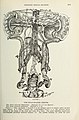
 Absorbing vessels of the human body, by W. Cruikshank Wellcome L0013059.jpg 3,683 × 4,744; 5.26 MB
-
 ACDF diagram1.png 3,256 × 1,394; 384 KB
-
 An écorché in a classical landscape. Drawing by A. Joron. Wellcome L0027181.jpg 4,858 × 6,488; 9.26 MB
-
 Anathomia. 53v 'Wound man'; flesh tinted; weapons coloured. Wellcome L0045155.jpg 2,672 × 3,424; 2.65 MB
-
 Anatomical fugitive sheets Wellcome L0055100.jpg 4,386 × 6,630; 8.8 MB
-
 Anatomical illustration showing female reproductive organs Wellcome L0047392.jpg 2,664 × 3,422; 1.96 MB
-
 Anatomical study of the head of Henry Jenkins, aged 169. Pen Wellcome V0007149.jpg 2,350 × 3,007; 2.61 MB
-

-
 Anatomy of the anal canal.png 3,312 × 3,980; 2.24 MB
-
 Anatomy of the Human Vagina.jpg 2,674 × 1,944; 2.42 MB
-
 Ball Collection, Acc 18692 (3154791724).jpg 697 × 978; 400 KB
-
 Bartolommeo da Arezzo - Study of a Flayed Torso - 1975.26.b - Cleveland Museum of Art.jpg 2,328 × 3,400; 1.93 MB
-

-
 Bartolommeo da Arezzo - Two Studies of a Flayed Man - 1975.26.a - Cleveland Museum of Art.tif 5,343 × 7,735; 118.25 MB
-
 Battista Franco - Arm Bones - 1964.379 - Cleveland Museum of Art.tif 6,484 × 2,163; 40.14 MB
-
 Battista Franco - Half-Length Skeleton in Profile - 1964.380.1 - Cleveland Museum of Art.tif 3,663 × 5,264; 55.18 MB
-

-

-
 Battista Franco - Rib Cages - 1964.383 - Cleveland Museum of Art.tif 6,330 × 3,163; 57.3 MB
-
 Battista Franco - Skeleton - 1964.380 - Cleveland Museum of Art.tif 5,401 × 4,992; 77.15 MB
-
 Battista Franco - Torsos with Rib Cages - 1964.378 - Cleveland Museum of Art.tif 6,529 × 2,405; 44.94 MB
-

-
 Biotronicrestrict.jpg 214 × 352; 51 KB
-
 Biotronicrestrict2.jpg 214 × 352; 59 KB
-
 Birth of Eve in the Garden of Eden and anatomical figure. Wellcome L0007123.jpg 1,156 × 1,594; 719 KB
-
 Book Illustration, Art Anatomy, Pectoralis Major in Use, 1884 (CH 68775931).jpg 4,096 × 2,992; 10.45 MB
-
 Bowel Ostomies.jpg 720 × 405; 68 KB
-
 Bowel resection and temporary colostomy illustration.jpg 1,414 × 2,700; 806 KB
-
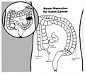 Bowel resection illustration.jpg 2,054 × 1,801; 489 KB
-
 Chinese manuscript Chiang-su Shang-hai Mo-hai shu-kuan. Wellcome L0020653.jpg 1,358 × 1,314; 803 KB
-
 Colon4.png 324 × 349; 14 KB
-
 Cpg291.jpg 651 × 941; 521 KB
-
 Desmoxytes (10.3897-zookeys.761.24214) Figure 3.jpg 1,378 × 1,847; 623 KB
-
 Drawing and CT Scan of Normal Epiglottis and Surrounding Structures.png 3,195 × 2,291; 8.77 MB
-

-
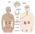 Endocrine Galego.svg 930 × 916; 394 KB
-

-
 EpiloicAppendices.png 600 × 503; 323 KB
-
 Experimental inquiries- part the third Fleuron T063463-5.png 1,451 × 1,094; 129 KB
-
 Eye illustration.webp 342 × 236; 23 KB
-

-
 Françoise Foliot - Jacques-Fabien Gautier-Dagoty - Structure du corps humain.jpg 3,626 × 5,368; 22.25 MB
-
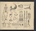
 Iconographic Encyclopedia of Science, Literature and Art 147.jpg 2,857 × 2,287; 939 KB
-
 Illustration of the heart, 17th century Wellcome L0002632.jpg 1,910 × 5,495; 3.55 MB
-
 Imada-Tsukanu-Domyaku Ichiranzu-1887.jpg 1,900 × 4,436; 5.67 MB
-
 Lateral lemniscus it.png 420 × 550; 33 KB
-
 Med template logo.png 516 × 893; 536 KB
-
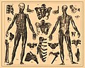
 Myologie de l'homme 1.jpg 2,388 × 3,312; 6.05 MB
-
 Myologie de l'homme 2.jpg 954 × 1,520; 797 KB
-
 OrbisPictus b 078.jpg 1,864 × 1,430; 1.14 MB
-
 OrbisPictus b 080.jpg 1,836 × 1,406; 995 KB
-
 OrbisPictus b 082.jpg 1,824 × 1,418; 556 KB
-
 OrbisPictus b 084.jpg 1,870 × 1,432; 561 KB
-
 OrbisPictus b 086.jpg 1,862 × 1,438; 923 KB
-
 Organ of corti it.svg 1,251 × 1,001; 104 KB
-
 Ottův slovník naučný - obrázek č. 0995.jpg 653 × 1,518; 577 KB
-

-
 Plate 3 - on stone by Jas. Queen ; P.S. Duval Lith. Phila. LCCN2014649311.jpg 4,112 × 5,120; 4.02 MB
-

-
 Quarta musculos commonstrantium figura, omnes in posteriori corporis LCCN92517416.jpg 1,071 × 1,536; 233 KB
-
 Rectal exam illustration.jpg 1,800 × 2,170; 602 KB
-
 Reisch, Margarita philosophica, 1503 Wellcome L0030314.jpg 1,186 × 1,730; 420 KB
-
 Right ventricle interior, circa 1749. Wellcome L0000262.jpg 2,354 × 1,890; 1.53 MB
-
 Sympathoadrenal System.jpg 736 × 591; 97 KB
-
 Tavola Mascagni.jpg 2,074 × 4,724; 516 KB
-
 Temporal bone - The Hungry Artist Multimedia.jpg 2,500 × 2,500; 1.54 MB
-

-
 The Ayurvedic Man., c.18th century Wellcome L0017592.jpg 2,686 × 4,151; 5.56 MB
-
 The heart, circa 1749. Wellcome L0004130.jpg 1,592 × 1,198; 977 KB
-

-

-

-
-

-

-

-

-
 Voie de Moore.jpg 255 × 134; 25 KB
-
 Waterston 1905 Heart and Pericardium Number 9 b21271252 001 0028 (cropped).jpg 2,205 × 1,246; 236 KB
-
 Waterston 1905 Heart and Pericardium Number 9 b21271252 001 0028.jpg 2,519 × 3,186; 832 KB
-
 Wellcome Chinese Collection 2 Wellcome L0067180.jpg 4,887 × 7,816; 12.21 MB
-
 Wisconsin medical recorder (1909) (14763058072).jpg 2,965 × 4,502; 1.56 MB
-
 Xiyuanlu jizheng-1843-Bones.jpg 2,800 × 2,362; 6.68 MB
-
 Yamawaki-Toyo-Zoshi-9-Organs.jpg 372 × 567; 227 KB
-
 Atlas historji naturalnej 1900 (118207844).jpg 5,003 × 4,268; 3.94 MB
-
 Atlas historji naturalnej 1900 (118207849).jpg 5,120 × 4,340; 3.95 MB
-
 Atlas historji naturalnej 1900 (118207858).jpg 5,003 × 4,268; 4.39 MB
-
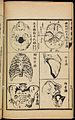
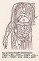 মানবদেহের অভ্যন্তরীণ অঙ্গপ্রত্যঙ্গের চিত্র.jpg 687 × 1,057; 257 KB

.jpg)


.jpg)

_Study_of_a_Flayed_Torso_(verso)_-_1975.26_-_Cleveland_Museum_of_Art.jpg)


.jpg)


_Figure_3.jpg)

.jpg)


_(14577610268).jpg)


_(14584365160).jpg)
_(14770755072).jpg)
_(14598085180).jpg)
_(14781656971).jpg)
_(14782448724).jpg)
_(14784448062).jpg)
_(14780810704).jpg)

.jpg)


_(14763058072).jpg)


.jpg)
.jpg)
.jpg)

_(14781657661).jpg)