Category:Histopathology of the female reproductive system
Jump to navigation
Jump to search
Subcategories
This category has the following 9 subcategories, out of 9 total.
F
H
N
O
P
U
V
Pages in category "Histopathology of the female reproductive system"
This category contains only the following page.
Media in category "Histopathology of the female reproductive system"
The following 151 files are in this category, out of 151 total.
-
Adenomatoid tumour - high mag.jpg 4,272 × 2,848; 5.65 MB
-
Adenomatoid tumour - intermed mag.jpg 4,272 × 2,848; 5.78 MB
-
Adenomatoid tumour - low mag.jpg 4,272 × 2,848; 5.39 MB
-
Adenomatoid tumour - very low mag.jpg 3,828 × 2,546; 4.92 MB
-
Adenomatoid tumour -a- very high mag.jpg 4,272 × 2,848; 5.86 MB
-
Adenomatoid tumour -b- very high mag.jpg 4,272 × 2,848; 5.78 MB
-
Atypical leiomyoma high mag.jpg 2,848 × 4,272; 5.5 MB
-
Atypical leiomyoma intermed mag.jpg 2,848 × 4,272; 4.79 MB
-
Atypical leiomyoma low mag.jpg 2,848 × 3,020; 1.23 MB
-
Atypical polypoid adenomyoma - add - intermed mag.jpg 2,848 × 4,272; 4.86 MB
-
Atypical polypoid adenomyoma - high mag.jpg 2,848 × 4,272; 5.54 MB
-
Atypical polypoid adenomyoma - intermed mag.jpg 4,272 × 2,848; 5.2 MB
-
Atypical polypoid adenomyoma - very high mag.jpg 2,848 × 4,272; 4.68 MB
-
Carcinosarcoma - 2 - high mag.jpg 4,272 × 2,848; 6.52 MB
-
Carcinosarcoma - 2 - intermed mag.jpg 4,272 × 2,848; 7.38 MB
-
Carcinosarcoma - 2 - very high mag.jpg 4,272 × 2,848; 4.89 MB
-
Carcinosarcoma - high mag.jpg 4,272 × 2,848; 5.77 MB
-
Carcinosarcoma - intermed mag.jpg 4,272 × 2,848; 6.71 MB
-
Carcinosarcoma - low mag.jpg 4,272 × 2,848; 6.91 MB
-
Carcinosarcoma - very high mag.jpg 4,272 × 2,848; 4.41 MB
-
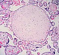
Cartilaginous metaplasia in placenta (?) 1 (590387907).jpg 1,470 × 1,375; 638 KB
-
Chorioamnionitis1.jpg 2,048 × 1,536; 746 KB
-
Chorioamnionitis2.jpg 2,048 × 1,536; 963 KB
-
Choroid plexus in teratoma -- high mag.jpg 4,272 × 2,848; 5.31 MB
-
Choroid plexus in teratoma -- intermed mag.jpg 4,272 × 2,848; 5.24 MB
-
Choroid plexus in teratoma -- low mag.jpg 4,272 × 2,848; 5.31 MB
-
Clear cell carcinoma - gynecologic tract - alt -- very high mag.jpg 4,272 × 2,848; 5.1 MB
-
Clear cell carcinoma - gynecologic tract - HE -- intermed mag.jpg 4,272 × 2,848; 5.93 MB
-
Clear cell carcinoma - gynecologic tract - PAS -- high mag.jpg 4,272 × 2,848; 4.71 MB
-
Clear cell carcinoma - gynecologic tract - PAS -- intermed mag.jpg 4,272 × 2,848; 4.61 MB
-
Clear cell carcinoma - gynecologic tract - PASD -- high mag.jpg 4,272 × 2,848; 4.66 MB
-
Clear cell carcinoma - gynecologic tract - PASD -- intermed mag.jpg 4,272 × 2,848; 4.13 MB
-
Clear cell carcinoma - gynecologic tract - PASD -- very high mag.jpg 4,272 × 2,848; 4.25 MB
-
Clear cell carcinoma - gynecologic tract -- high mag.jpg 4,272 × 2,848; 5.09 MB
-
Clear cell carcinoma - gynecologic tract -- intermed mag.jpg 4,272 × 2,848; 5.12 MB
-
Clear cell carcinoma - gynecologic tract -- low mag.jpg 4,272 × 2,848; 8.3 MB
-
Clear cell carcinoma - gynecologic tract -- very high mag.jpg 4,272 × 2,848; 4.94 MB
-
Condyloma acuminatum - high mag.jpg 4,272 × 2,848; 4.33 MB
-
Condyloma acuminatum - intermed mag.jpg 4,272 × 2,848; 4.53 MB
-
Condyloma acuminatum - low mag.jpg 4,272 × 2,848; 4.65 MB
-
Condyloma acuminatum - very high mag.jpg 4,272 × 2,848; 4.47 MB
-
Dermatomycosis - gms - high mag.jpg 4,272 × 2,848; 3.72 MB
-
Dermatomycosis - gms - intermed mag.jpg 4,272 × 2,848; 3.75 MB
-
Dermatomycosis - gms - low mag.jpg 4,272 × 2,848; 4.18 MB
-
Dermatomycosis - gms - very high mag.jpg 4,272 × 2,848; 3.78 MB
-
Dermatomycosis - high mag.jpg 2,848 × 4,272; 5.18 MB
-
Dermatomycosis - intermed mag.jpg 2,848 × 4,272; 6.7 MB
-
Dermatomycosis - low mag.jpg 4,272 × 2,848; 5.34 MB
-
Differentiated vulvar intraepithelial neoplasia - deep - high mag.jpg 2,848 × 4,272; 5.95 MB
-
Differentiated vulvar intraepithelial neoplasia - intermed mag.jpg 2,848 × 4,272; 5.05 MB
-
Differentiated vulvar intraepithelial neoplasia - low mag.jpg 4,272 × 2,848; 4.26 MB
-
Differentiated vulvar intraepithelial neoplasia - superficial - high mag.jpg 2,848 × 4,272; 4.98 MB
-
Ectopic pregnancy - decidua -- intermed mag.jpg 4,272 × 2,848; 6.56 MB
-
Ectopic pregnancy -- high mag.jpg 4,272 × 2,848; 5.66 MB
-
Ectopic pregnancy -- intermed mag.jpg 4,272 × 2,848; 5.48 MB
-
Ectopic pregnancy -- low mag.jpg 4,272 × 2,848; 6.74 MB
-
Endometrium with SPRM changes - a -- extremely low mag.jpg 6,000 × 4,000; 11.33 MB
-
Endometrium with SPRM changes - a -- very low mag.jpg 6,000 × 4,000; 11.99 MB
-
Endometrium with SPRM changes - b -- low mag.jpg 4,000 × 6,000; 11.29 MB
-
Endometrium with SPRM changes - b -- very low mag.jpg 6,000 × 4,000; 11.94 MB
-
Endometrium with SPRM changes - c -- high mag.jpg 4,000 × 6,000; 9.71 MB
-
Endometrium with SPRM changes - c -- low mag.jpg 6,000 × 4,000; 10.71 MB
-
Endometrium with SPRM changes - c -- very low mag.jpg 6,000 × 4,000; 10.8 MB
-
Endometrium with SPRM changes - c1 -- intermed mag.jpg 6,000 × 4,000; 10.92 MB
-
Endometrium with SPRM changes - c1 -- very high mag.jpg 4,000 × 6,000; 8.28 MB
-
Endometrium with SPRM changes - c2 -- intermed mag.jpg 4,000 × 6,000; 9.99 MB
-
Endometrium with SPRM changes - c2 -- very high mag.jpg 4,000 × 6,000; 8.13 MB
-
Exocervix -- extremely high mag.jpg 4,272 × 2,848; 5.68 MB
-
Exocervix -- high mag.jpg 4,272 × 2,848; 7.43 MB
-
Exocervix -- very high mag.jpg 4,272 × 2,848; 6.34 MB
-
Female adnexal tumour of probable Wolffian origin - high mag.jpg 2,848 × 4,272; 6.39 MB
-
Female adnexal tumour of probable Wolffian origin - intermed mag.jpg 2,848 × 4,272; 7.67 MB
-
Female adnexal tumour of probable Wolffian origin - very high mag.jpg 2,848 × 4,272; 6.34 MB
-
Fetal intermediate cellular type rhabdomyoma.JPG 550 × 550; 102 KB
-
Fibroepithelial polyp of the vulva with atypical cells on low magnification.jpg 2,592 × 1,944; 788 KB
-
Glassy cell carcinoma - high mag.jpg 4,272 × 2,848; 4.98 MB
-
Glassy cell carcinoma - intermed mag.jpg 4,272 × 2,848; 5.6 MB
-
Glassy cell carcinoma - low mag.jpg 4,272 × 2,848; 5.57 MB
-
Glassy cell carcinoma - very high mag.jpg 4,272 × 2,848; 4.37 MB
-
High grade squamous intraepithelial lesion - 2 - ki67 -- high mag.jpg 4,272 × 2,848; 4.44 MB
-
High grade squamous intraepithelial lesion - 2 - ki67 -- intermed mag.jpg 4,272 × 2,848; 4.84 MB
-
High grade squamous intraepithelial lesion - 2 - p16 -- high mag.jpg 4,272 × 2,848; 4.37 MB
-
High grade squamous intraepithelial lesion - 2 - p16 -- intermed mag.jpg 4,272 × 2,848; 4.23 MB
-
High grade squamous intraepithelial lesion - 2 - p63 -- high mag.jpg 4,272 × 2,848; 4.46 MB
-
High grade squamous intraepithelial lesion - 2 - p63 -- intermed mag.jpg 4,272 × 2,848; 4.37 MB
-
High grade squamous intraepithelial lesion - 2 -- high mag.jpg 4,272 × 2,848; 5.5 MB
-
High grade squamous intraepithelial lesion - 2 -- intermed mag.jpg 4,272 × 2,848; 5.97 MB
-
High grade squamous intraepithelial lesion - 2 -- very high mag.jpg 2,848 × 4,272; 5.37 MB
-
High-grade sqaumous intraepithelial lesion -- high mag.jpg 2,848 × 4,272; 5.37 MB
-
High-grade squamous intraepithelial lesion - p16 -- high mag.jpg 4,272 × 2,848; 4.53 MB
-
High-grade squamous intraepithelial lesion -- intermed mag.jpg 4,272 × 2,848; 4.12 MB
-
Histological sections of ovarian cortex from patients with TS.png 722 × 336; 602 KB
-
Intermediate magnification of a borderline serous tumor showing eosiniophilic cells.jpg 2,592 × 1,944; 914 KB
-
Keratinized cervix -- high mag.jpg 4,272 × 2,848; 5.23 MB
-
Keratinized cervix -- intermed mag.jpg 4,272 × 2,848; 5.91 MB
-
Keratinized cervix -- very high mag.jpg 2,848 × 4,272; 5.71 MB
-
Lipoleiomyoma1.jpg 2,048 × 1,536; 895 KB
-
Lipoleiomyoma2.jpg 2,048 × 1,536; 835 KB
-
Luteinized follicular cyst.jpg 2,469 × 2,848; 3.16 MB
-
Microglandular hyperplasia - alt 1 -- high mag.jpg 4,272 × 2,848; 5.86 MB
-
Microglandular hyperplasia - alt 2 -- high mag.jpg 4,272 × 2,848; 5.25 MB
-
Microglandular hyperplasia -- high mag.jpg 4,272 × 2,848; 5.76 MB
-
Microglandular hyperplasia -- intermed mag.jpg 4,272 × 2,848; 5.83 MB
-
Microglandular hyperplasia -- low mag.jpg 4,272 × 2,848; 5.55 MB
-
Muellerian cyst - 2 -- high mag.jpg 4,272 × 2,848; 4.5 MB
-
Muellerian cyst - 2 -- intermed mag.jpg 4,272 × 2,848; 4.9 MB
-
Muellerian cyst -- high mag.jpg 4,272 × 2,848; 3.97 MB
-
Muellerian cyst -- intermed mag.jpg 4,272 × 2,848; 4.23 MB
-
Muellerian cyst -- very high mag.jpg 4,272 × 2,848; 4.42 MB
-
Papillary hidradenoma - high mag.jpg 2,848 × 4,272; 4.9 MB
-
Papillary hidradenoma - intermed mag.jpg 2,848 × 4,272; 4.58 MB
-
Papillary hidradenoma - very high mag.jpg 2,848 × 4,272; 3.59 MB
-
Plasmacytosis mucosae - alt -- high mag.jpg 4,272 × 2,848; 5.4 MB
-
Plasmacytosis mucosae -- high mag.jpg 4,272 × 2,848; 5.23 MB
-
Plasmacytosis mucosae -- intermed mag.jpg 4,272 × 2,848; 5.67 MB
-
Plasmacytosis mucosae -- very high mag.jpg 4,272 × 2,848; 4.39 MB
-
Products of conception - intermed mag.jpg 4,272 × 2,848; 4.52 MB
-
Secretory phase endometrium -- high mag.jpg 2,848 × 4,272; 4.94 MB
-
Secretory phase endometrium -- intermed mag.jpg 2,848 × 4,272; 5.74 MB
-
Secretory phase endometrium -- low mag.jpg 4,272 × 2,848; 6.57 MB
-
Serous borderline tumour with micropapillary pattern - a -- high mag.jpg 2,848 × 4,272; 4.94 MB
-
Serous borderline tumour with micropapillary pattern - a -- intermed mag.jpg 2,848 × 4,272; 5.27 MB
-
Serous borderline tumour with micropapillary pattern - a -- low mag.jpg 2,848 × 4,272; 4.83 MB
-
Serous borderline tumour with micropapillary pattern - a -- very high mag.jpg 4,272 × 2,848; 3.81 MB
-
Serous borderline tumour with micropapillary pattern - a -- very low mag.jpg 4,272 × 2,848; 4.52 MB
-
Serous borderline tumour with micropapillary pattern - a2 -- high mag.jpg 4,272 × 2,848; 4.53 MB
-
Serous borderline tumour with micropapillary pattern - a3 -- high mag.jpg 4,272 × 2,848; 4.67 MB
-
Serous borderline tumour with micropapillary pattern -- high mag.jpg 4,272 × 2,848; 4.17 MB
-
Serous borderline tumour with micropapillary pattern -- intermed mag.jpg 4,272 × 2,848; 4.97 MB
-
Serous borderline tumour with micropapillary pattern -- low mag.jpg 4,272 × 2,848; 5.23 MB
-
Serous borderline tumour with micropapillary pattern -- very low mag.jpg 4,272 × 2,848; 4.48 MB
-
Serous borderline tumour with micropapillary pattern -- very very low mag.jpg 4,272 × 2,848; 4.08 MB
-
Serous cystadenofibroma -- high mag.jpg 4,272 × 2,848; 5.37 MB
-
Serous cystadenofibroma -- intermed mag.jpg 4,272 × 2,848; 5.63 MB
-
Serous cystadenofibroma -- low mag.jpg 4,272 × 2,848; 4.65 MB
-
Serous cystadenofibroma -- very low mag.jpg 4,272 × 2,848; 5.57 MB
-
Simple endometrial hyperplasia - high mag.jpg 2,848 × 4,272; 5.99 MB
-
Simple endometrial hyperplasia - intermed mag.jpg 2,848 × 4,272; 5.51 MB
-
Simple endometrial hyperplasia - low mag.jpg 3,952 × 2,784; 5.01 MB
-
Strumal carcinoid - high mag.jpg 4,272 × 2,848; 6.05 MB
-
Strumal carcinoid - intermed mag.jpg 4,272 × 2,848; 7.42 MB
-
Strumal carcinoid - very high mag.jpg 4,272 × 2,848; 6.2 MB
-
Tunnel cluster - high mag.jpg 4,272 × 2,848; 5.1 MB
-
Tunnel cluster - intermed mag.jpg 4,272 × 2,848; 5.63 MB
-
Tunnel cluster - low mag.jpg 4,272 × 2,848; 5.26 MB
-
Tunnel cluster - very high mag.jpg 4,272 × 2,848; 4.11 MB
-
Tunnel cluster - very low mag.jpg 4,272 × 2,848; 6.32 MB
-
Vulvar intraepithelial neoplasia3 1.jpg 1,281 × 1,746; 878 KB
-
Vulvar intraepithelial neoplasia3 2.jpg 1,536 × 1,257; 668 KB
-
Xanthomatous endocervical polyp -- intermed mag.jpg 4,272 × 2,848; 4.27 MB
-
Xanthomatous endocervical polyp -- low mag.jpg 4,272 × 2,848; 3.55 MB


