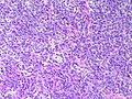
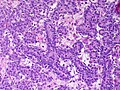
Category:Histopathology of ovarian Sertoli-Leydig cell tumor
Jump to navigation
Jump to search
Media in category "Histopathology of ovarian Sertoli-Leydig cell tumor"
The following 16 files are in this category, out of 16 total.
-
 Ovary SertoliLeydig Intermediate HP CTR.jpg 2,048 × 1,536; 1.2 MB
-
 Ovary SertoliLeydig Intermediate HP2 CTR.jpg 2,048 × 1,536; 1.48 MB
-
 Ovary SertoliLeydig Intermediate MP CTR.jpg 2,048 × 1,536; 1.53 MB
-
 Ovary SertoliLeydigCellTumor 3 PA.jpg 2,048 × 1,536; 2.48 MB
-
 Ovary SertoliLeydigCellTumor 4 PA.jpg 2,048 × 1,536; 2.87 MB
-
 Ovary SertoliLeydigCellTumor 5 PA.jpg 2,048 × 1,536; 2.87 MB
-
 Ovary SertoliLeydigCellTumor 6 PA.jpg 2,048 × 1,536; 3.04 MB
-
 Ovary SertoliLeydigCellTumor 7 PA.jpg 2,048 × 1,536; 3.23 MB
-
 Ovary SertoliLeydigCellTumor HP PA.JPG 2,301 × 3,070; 1.4 MB
-
 Ovary SertoliLeydigCellTumor LP PA.JPG 3,070 × 2,301; 1.46 MB
-
 Ovary SertoliLeydigCellTumor MP CTR.jpg 2,048 × 1,536; 1.15 MB
-
 Ovary SertoliLeydigCellTumor MP PA.JPG 3,070 × 2,301; 1.81 MB
-
 Ovary SertoliLeydigCellTumor MP2 CTR.jpg 2,048 × 1,536; 2.38 MB
-
 Ovary SertoliLeydigCellTumor MP3 CTR.jpg 2,048 × 1,536; 2.07 MB
-
 Ovary SertoliLeydigCellTumor PA.jpg 2,048 × 1,536; 2.68 MB
-
 Ovary SertoliLeydigCellTumor.jpg 2,048 × 1,536; 2.52 MB


