Category:Histopathology of colorectal adenocarcinoma
Jump to navigation
Jump to search
Subcategories
This category has only the following subcategory.
Media in category "Histopathology of colorectal adenocarcinoma"
The following 95 files are in this category, out of 95 total.
-
 Adenocarcinoma coli.jpg 640 × 512; 77 KB
-
 Adenocarcinoma of the colon-histology.JPG 3,072 × 2,304; 2.58 MB
-
 Cecal adenocarcinoma.jpg 2,048 × 1,536; 661 KB
-
 Colon adenocarcinoma - biopsy, high mag.jpg 4,272 × 2,848; 5.56 MB
-
 Colon adenocarcinoma - biopsy, intermed. mag.1.jpg 4,272 × 2,848; 6.78 MB
-
 Colon adenocarcinoma - biopsy, intermed. mag.2.jpg 4,272 × 2,848; 4.42 MB
-
 Colon adenocarcinoma - biopsy, low mag.jpg 4,272 × 2,848; 2.43 MB
-
 Colon adenocarcinoma - biopsy, very high mag.jpg 4,272 × 2,848; 4.78 MB
-
 Colon Adenocarcinoma, Whole-Mount Scan (1858505117).jpg 3,639 × 2,426; 6.58 MB
-
 Colonic adenocarcinoma (1) Endoscopic biosy.jpg 700 × 527; 175 KB
-
 Colonic Adenocarcinoma ex Villous Adenoma.jpg 2,082 × 2,223; 1.87 MB
-
 Colonic mucinous adenocarcinoma - low mag.jpg 2,848 × 4,272; 4.72 MB
-
 Colonic mucinous adenocarcinoma - very low mag.jpg 4,272 × 2,848; 5.29 MB
-
 Colorectal adenocarcinoma - alt -- high mag.jpg 4,272 × 2,848; 5.71 MB
-
 Colorectal adenocarcinoma - alt -- intermed mag.jpg 4,272 × 2,848; 5.82 MB
-
 Colorectal adenocarcinoma - alt -- low mag.jpg 4,272 × 2,848; 6.22 MB
-
 Colorectal adenocarcinoma - cribriform pattern, high mag.jpg 4,272 × 2,848; 5.31 MB
-
 Colorectal adenocarcinoma - cribriform pattern, intermed. mag.jpg 4,272 × 2,848; 6.26 MB
-
 Colorectal adenocarcinoma - cribriform pattern, low mag.jpg 4,272 × 2,848; 7 MB
-
 Colorectal adenocarcinoma - cribriform pattern, very high mag.jpg 4,272 × 2,848; 4.64 MB
-
 Colorectal adenocarcinoma - pT2, high mag.jpg 4,272 × 2,848; 5.57 MB
-
 Colorectal adenocarcinoma - pT2, intermed. mag.jpg 4,272 × 2,848; 6.36 MB
-
 Colorectal adenocarcinoma - pT2, low mag.jpg 4,272 × 2,848; 6.55 MB
-
 Colorectal adenocarcinoma - pT2, very high mag.jpg 4,272 × 2,848; 4.8 MB
-
 Colorectal adenocarcinoma - pT3, high mag.1.jpg 4,272 × 2,848; 4.63 MB
-
 Colorectal adenocarcinoma - pT3, high mag.2.jpg 4,272 × 2,848; 4.1 MB
-
 Colorectal adenocarcinoma - pT3, intermed. mag.1.jpg 4,272 × 2,848; 4.4 MB
-
 Colorectal adenocarcinoma - pT3, intermed. mag.2.jpg 4,272 × 2,848; 4.84 MB
-
 Colorectal adenocarcinoma - pT3, low mag.jpg 4,272 × 2,848; 4.75 MB
-
 Colorectal adenocarcinoma - pT3, very high mag.jpg 4,272 × 2,848; 4.32 MB
-
 Colorectal adenocarcinoma - pT4a, high mag.1.jpg 4,272 × 2,848; 3.68 MB
-
 Colorectal adenocarcinoma - pT4a, high mag.2.jpg 4,272 × 2,848; 4.22 MB
-
 Colorectal adenocarcinoma - pT4a, intermed. mag.jpg 4,272 × 2,848; 4.36 MB
-
 Colorectal adenocarcinoma - pT4a, low mag.jpg 4,272 × 2,848; 4.45 MB
-
 Colorectal adenocarcinoma - pT4a, very high mag.1.jpg 4,272 × 2,848; 3.72 MB
-
 Colorectal adenocarcinoma - pT4a, very high mag.2.jpg 4,272 × 2,848; 4.76 MB
-
 Colorectal adenocarcinoma - solid pattern, high mag.jpg 4,272 × 2,848; 6.21 MB
-
 Colorectal adenocarcinoma - solid pattern, intermed. mag.jpg 4,272 × 2,848; 7.64 MB
-
 Colorectal adenocarcinoma - solid pattern, low mag.jpg 4,272 × 2,848; 6.36 MB
-
 Colorectal adenocarcinoma - solid pattern, very high mag.jpg 4,272 × 2,848; 5.1 MB
-
 Colorectal adenocarcinoma -- high mag.jpg 4,272 × 2,848; 5.51 MB
-
 Colorectal adenocarcinoma -- intermed mag.jpg 4,272 × 2,848; 5.59 MB
-
 Colorectal adenocarcinoma -- low mag.jpg 4,272 × 2,848; 6.14 MB
-
 Colorectal adenocarcinoma with MMR - MLH1 -- high mag.jpg 6,000 × 4,000; 8.14 MB
-
 Colorectal adenocarcinoma with MMR - MLH1 -- intermed mag.jpg 6,000 × 4,000; 9.56 MB
-
 Colorectal adenocarcinoma with MMR - MSH2 -- high mag.jpg 6,000 × 4,000; 8.42 MB
-
 Colorectal adenocarcinoma with MMR - MSH2 -- intermed mag.jpg 6,000 × 4,000; 8.45 MB
-
 Colorectal adenocarcinoma with MMR - MSH6 -- high mag.jpg 6,000 × 4,000; 8.35 MB
-
 Colorectal adenocarcinoma with MMR - MSH6 -- intermed mag.jpg 6,000 × 4,000; 9.55 MB
-
 Colorectal adenocarcinoma with MMR - PMS2 -- high mag.jpg 6,000 × 4,000; 8.49 MB
-
 Colorectal adenocarcinoma with MMR - PMS2 -- intermed mag.jpg 6,000 × 4,000; 9.36 MB
-
 Colorectal adenocarcinoma with MMR -- high mag.jpg 6,000 × 4,000; 8.75 MB
-
 Colorectal adenocarcinoma with MMR -- intermed mag.jpg 6,000 × 4,000; 9.99 MB
-
 Colorectal adenocarcinoma with MMR -- low mag.jpg 6,000 × 4,000; 11.44 MB
-
 Colorectal adenocarcinoma with mucinous differentiation, high mag.jpg 4,272 × 2,848; 5.98 MB
-
 Colorectal adenocarcinoma with mucinous differentiation, intermed. mag.jpg 4,272 × 2,848; 2.66 MB
-
 Colorectal adenocarcinoma with mucinous differentiation, low mag.jpg 4,272 × 2,848; 6.36 MB
-
 Colorectal adenocarcinoma with mucinous differentiation, very high mag.jpg 4,272 × 2,848; 5.15 MB
-
 Colorectal adenocarcinoma, not otherwise specified.jpg 1,705 × 1,781; 1.87 MB
-
 Low-grade colorectal adenocarcinoma, high mag.jpg 4,272 × 2,848; 6.35 MB
-
 Low-grade colorectal adenocarcinoma, intermed. mag.jpg 4,272 × 2,848; 7.63 MB
-
 Low-grade colorectal adenocarcinoma, low mag.jpg 4,272 × 2,848; 7 MB
-
 Low-grade colorectal adenocarcinoma, very high mag.jpg 4,272 × 2,848; 5.15 MB
-
 Lymph node with metastatic adenocarcinoma, high mag.jpg 4,272 × 2,848; 5.83 MB
-
 Lymph node with metastatic adenocarcinoma, intermed. mag.1.jpg 4,272 × 2,848; 6.51 MB
-
 Lymph node with metastatic adenocarcinoma, intermed. mag.2.jpg 4,272 × 2,848; 6.52 MB
-
 Lymph node with metastatic adenocarcinoma, low mag.jpg 4,272 × 2,848; 7.48 MB
-
 Lymph node with metastatic adenocarcinoma, very high mag.jpg 4,272 × 2,848; 5.04 MB
-
 Metastatic adenocarcinoma - cerebellum - very low mag.jpg 4,272 × 2,848; 6.7 MB
-
 Metastatic colonic adenocarcinoma - Case 263 (8558729323).jpg 3,264 × 2,448; 3.06 MB
-
 Metastatic colonic adenocarcinoma - Case 263 (8558729543).jpg 3,264 × 2,448; 2.2 MB
-
 Metastatic Colonic Adenocarcinoma in Lymph Node.jpg 1,004 × 1,031; 268 KB
-
 Metastatic colonic adenocarcinoma, intrabronchial (7425443926).jpg 2,560 × 1,920; 2.62 MB
-
 MSI-H colorectal adenocarcinoma - MLH1.jpg 4,272 × 2,848; 4.91 MB
-
 MSI-H colorectal adenocarcinoma - MSH2.jpg 4,272 × 2,848; 4.33 MB
-
 MSI-H colorectal adenocarcinoma - MSH6.jpg 4,272 × 2,848; 4.42 MB
-
 MSI-H colorectal adenocarcinoma - PMS2.jpg 4,272 × 2,848; 4.83 MB
-
 Mucinous adenocarcinoma of the colon, HE 1.JPG 1,920 × 1,280; 1.43 MB
-
 Mucinous adenocarcinoma of the colon, HE 2.JPG 1,920 × 1,280; 1.25 MB
-
 Mucinous adenocarcinoma of the colon, HE 3.JPG 1,920 × 1,280; 1.18 MB
-
 Mucinous adenocarcinoma of the colon, HE 4.JPG 1,920 × 1,280; 1.32 MB
-
 Mucinous adenocarcinoma of the colon, HE 5.JPG 1,920 × 1,280; 1.68 MB
-
 Mucinous colorectal adenocarcinoma, high mag.jpg 4,272 × 2,848; 1.58 MB
-
 Mucinous colorectal adenocarcinoma, intermed. mag.1.jpg 4,272 × 2,848; 2.04 MB
-
 Mucinous colorectal adenocarcinoma, intermed. mag.2.jpg 4,272 × 2,848; 4.98 MB
-
 Mucinous colorectal adenocarcinoma, low mag.jpg 4,272 × 2,848; 2.48 MB
-
 Mucinous colorectal adenocarcinoma, very high mag.jpg 4,272 × 2,848; 1.23 MB
-
 Paracolonic Lymph Node with Metastatic Adenocarcinoma (6796280527).jpg 1,581 × 1,051; 486 KB
-
 Paracolonic Lymph Node with Metastatic Adenocarcinoma (6796280631).jpg 1,597 × 1,063; 524 KB
-
 Positive margin with cautery artefact - adenocarcinoma - high mag.jpg 2,848 × 4,272; 4.82 MB
-
 Positive margin with cautery artefact - adenocarcinoma - intermed mag.jpg 2,848 × 4,272; 5.76 MB
-
 Positive margin with cautery artefact - adenocarcinoma - low mag.jpg 2,848 × 4,272; 5.57 MB
-
 Signet ring cell, original.jpg 1,029 × 855; 121 KB
-
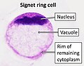 Signet ring cell.jpg 911 × 710; 115 KB
-
 Signet Ring Cells (2202231656).jpg 1,600 × 1,200; 263 KB


.jpg)
_Endoscopic_biosy.jpg)


.jpg)
.jpg)

.jpg)


.jpg)
.jpg)


.jpg)