Category:Histopathology of Rosai–Dorfman disease
Jump to navigation
Jump to search
Media in category "Histopathology of Rosai–Dorfman disease"
The following 19 files are in this category, out of 19 total.
-
 Emperipolesis - very high mag.jpg 1,551 × 1,204; 983 KB
-
 Emperipolesis rosai dorfman erkrankung.jpg 2,080 × 1,542; 773 KB
-
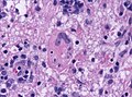
 Granuloma rosai dorfman brain.jpg 2,080 × 1,542; 969 KB
-
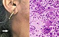
 Rosai dorfman disease.jpg 1,280 × 720; 79 KB
-
 Rosai-Dorfman disease - very high mag.jpg 4,272 × 2,848; 6.06 MB
-
-
 Rosai-Dorfman disease of parotid gland - alt -- high mag.jpg 4,000 × 6,000; 10.78 MB
-
 Rosai-Dorfman disease of parotid gland -- high mag.jpg 4,000 × 6,000; 16.71 MB
-
 Rosai-Dorfman disease of parotid gland -- intermed mag.jpg 4,000 × 6,000; 14.46 MB
-
 Rosai-Dorfman disease of parotid gland -- low mag.jpg 4,000 × 6,000; 11.11 MB
-
 Rosai-Dorfman disease of parotid gland -- very high mag.jpg 4,000 × 6,000; 11.06 MB
-
 Rosai-dorfman.jpg 465 × 512; 218 KB
-
 Sinus histiocytosis - deep -- high mag.jpg 2,848 × 4,272; 6.19 MB
-
 Sinus histiocytosis -- high mag.jpg 2,848 × 4,272; 6.31 MB
-
 Sinus histiocytosis -- intermed mag.jpg 2,848 × 4,272; 6.81 MB
-
 Sinushistiocytosis, HE 1.JPG 1,920 × 1,280; 1.87 MB
-
 Sinushistiocytosis, HE 2.JPG 1,920 × 1,280; 1.65 MB
-
 Sinushistiocytosis, HE 3.JPG 1,920 × 1,280; 1.33 MB
-
 Sinushistiocytosis, HE 4.JPG 1,920 × 1,280; 1.34 MB


