Category:Histology of the human pineal gland
Jump to navigation
Jump to search
Media in category "Histology of the human pineal gland"
The following 55 files are in this category, out of 55 total.
-
 Blood Vessels in Septae of Older Human Pineal Gland by Phase Contrast (46684388894).jpg 3,264 × 1,840; 1.57 MB
-
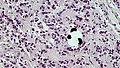
 Brain Sand in Older Human Pineal Gland (33527661718).jpg 3,264 × 1,840; 2.13 MB
-
 Brain Sand in Older Human Pineal Gland (40440641473).jpg 3,264 × 1,840; 5.03 MB
-
 Brain Sand in Older Human Pineal Gland (46491416155).jpg 3,264 × 1,840; 6.74 MB
-
 Brain Sand in Older Human Pineal Gland (47352928552).jpg 3,264 × 1,840; 1.91 MB
-
 Brain Sand in Older Human Pineal Gland (47353308772).jpg 3,264 × 1,840; 1.47 MB
-
 Brain Sand in Older Human Pineal Gland by Phase Contrast (46683386794).jpg 3,264 × 1,840; 2.48 MB
-
 Brain Sand in Older Human Pinealocyte by Phase Contrast (46491414525).jpg 3,264 × 1,840; 3.02 MB
-
 Brain Sand in Older Pineal Gland by Phase Contrast (46684389344).jpg 3,264 × 1,840; 1.91 MB
-
 Capillary Supply in Young Human Pineal Gland (40406904583).jpg 3,264 × 1,840; 2.21 MB
-

-
 Connective Tissue Wrapped Lobes in Older Human Pineal Gland (47352927622).jpg 3,264 × 1,840; 2.52 MB
-
 Connective Tissue Wrapped Lobes in Older PIneal Gland by Phase Contrast (46491415755).jpg 3,264 × 1,840; 3.02 MB
-
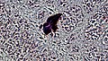
 Deposits of Sand in Older Human Pineal Gland by Phase Contrast (46683387694).jpg 3,264 × 1,840; 8.95 MB
-
 Human Pineal Gland Young (40406900493).jpg 3,264 × 1,840; 8.2 MB
-
 Human Pineal Gland Young (47319577772).jpg 3,264 × 1,840; 9.15 MB
-
 Human Pineal Gland Young (47372420061).jpg 3,264 × 1,840; 8.42 MB
-
 Human Pineal Gland Young by Phase Contrast (33496196008).jpg 3,264 × 1,840; 2.31 MB
-
 Human Pineal Gland Young by Phase Contrast (46457456425).jpg 3,264 × 1,840; 1.57 MB
-
 Human Pineal Gland Young by Phase Contrast (46457456745).jpg 3,264 × 1,840; 2.68 MB
-
 Human Pineal Gland Young by Phase Contrast (46457461585).jpg 3,264 × 1,840; 6.47 MB
-
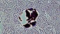
 Large Brain Sand Deposit in Older Human Pineal Gland (40442211613).jpg 3,264 × 1,840; 1.24 MB
-
 Large Deposit of Brain Sand in Older Human Pineal by Phase Contrast (47354891072).jpg 3,264 × 1,840; 5.31 MB
-
 Older Human Pituitary Gland by Phase Contrast (40442211443).jpg 3,264 × 1,840; 1.55 MB
-
 Pia Mater in Older Human Pineal Gland by Phase Contrast (40442210013).jpg 3,264 × 1,840; 2.16 MB
-
 Pineal gland - high mag.jpg 2,848 × 4,272; 6.35 MB
-
 Pineal gland - intermed mag.jpg 2,848 × 4,272; 6.91 MB
-
 Pineal gland - low mag.jpg 2,848 × 4,272; 7.99 MB
-
 Pineal gland - very high mag.jpg 2,848 × 4,272; 5.36 MB
-
 Pineal.jpg 4,080 × 3,072; 3.13 MB
-
 Pinealocyte Rosettes in Older Pineal Gland (40440642443).jpg 3,264 × 1,840; 2.1 MB
-
 Pinealocytes and Associated Astrocytes in Older Pineal Gland (33530193708).jpg 3,264 × 1,840; 1.61 MB
-
 Pinealocytes and Astrocytes in Young Human Pineal Gland (40406903583).jpg 3,264 × 1,840; 7.26 MB
-

-
 Rosette of Pinealocytes in Young Human Pineal Gland (40406903783).jpg 3,264 × 1,840; 8.12 MB
-
 Rosettes of Pinealocytes in Older Human Pineal Gland (47352928292).jpg 3,264 × 1,840; 2.54 MB
-
 Rosettes of Pinealocytes in Older Human Pineal Gland by Phase Contrast (40441156793).jpg 3,264 × 1,840; 3.04 MB
-
 Rosettes of Pinealocytes in Older Human Pineal Gland by Phase Contrast (47354890892).jpg 3,264 × 1,840; 1.58 MB
-
 Rosettes of Pinealocytes in Young Human Pineal Gland by Phase Contrast (46457461285).jpg 3,264 × 1,840; 6.11 MB
-
 Septate Spaces Between Lobes in Older Human Pineal Gland by Phase Contrast (40441155733).jpg 3,264 × 1,840; 3.06 MB
-
 Tightly Adherent Pia Mater in Older Human Pineal Gland (40440641833).jpg 3,264 × 1,840; 1.87 MB
-
 Tightly Adherent Pia Mater in Older Human Pineal Gland (47352927992).jpg 3,264 × 1,840; 1.99 MB
-
 Well Defined Lobes in Older Human Pineal Gland (33527661368).jpg 3,264 × 1,840; 2.6 MB
-
 Well Defined Lobes in Older Human Pineal Gland by Phase Contrast (40442209533).jpg 3,264 × 1,840; 1.43 MB
-
 Young Pineal Gland Hematoxylin and Eosin Staining (33496198538).jpg 3,264 × 1,840; 1.38 MB
-
 Young Pineal Gland Hematoxylin and Eosin Staining (33496198688).jpg 3,264 × 1,840; 1.36 MB
-
 Young Pineal Gland Hematoxylin and Eosin Staining (33496198818).jpg 3,264 × 1,840; 1.38 MB
-
 Young Pineal Gland Hematoxylin and Eosin Staining (33496198968).jpg 3,264 × 1,840; 1.41 MB
-
 Young Pineal Gland Hematoxylin and Eosin Staining (33496199108).jpg 3,264 × 1,840; 1.75 MB
-
 Young Pineal Gland Hematoxylin and Eosin Staining (46457459845).jpg 3,264 × 1,840; 2.15 MB
-
 Young Pineal Gland Hematoxylin and Eosin Staining (46457460095).jpg 3,264 × 1,840; 1.77 MB
-
 Young Pineal Gland Hematoxylin and Eosin Staining (46457460375).jpg 3,264 × 1,840; 1.71 MB
-
 Young Pineal Gland Hematoxylin and Eosin Staining (46457460555).jpg 3,264 × 1,840; 1.81 MB
-
 Young Pineal Gland Hematoxylin and Eosin Staining (46457460735).jpg 3,264 × 1,840; 1.77 MB
-
 Young Pineal Gland Hematoxylin and Eosin Staining (46457461065).jpg 3,264 × 1,840; 1.59 MB
.jpg)
.jpg)
.jpg)
.jpg)
.jpg)
.jpg)
.jpg)
.jpg)
.jpg)
.jpg)
.jpg)
.jpg)
.jpg)
.jpg)
.jpg)
.jpg)
.jpg)
.jpg)
.jpg)
.jpg)
.jpg)
.jpg)
.jpg)
.jpg)
.jpg)


.jpg)
.jpg)
.jpg)
.jpg)
.jpg)
.jpg)
.jpg)
.jpg)
.jpg)
.jpg)
.jpg)
.jpg)
.jpg)
.jpg)
.jpg)
.jpg)
.jpg)
.jpg)
.jpg)
.jpg)
.jpg)
.jpg)
.jpg)
.jpg)
.jpg)