Category:Histology of mammal skin
Jump to navigation
Jump to search
Subcategories
This category has the following 2 subcategories, out of 2 total.
H
Media in category "Histology of mammal skin"
The following 53 files are in this category, out of 53 total.
-
AchselhautHisto.jpeg 1,280 × 960; 92 KB
-
Basal lamina.jpg 347 × 109; 6 KB
-
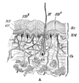
Cambridge Natural History Mammalia Fig 001a.png 494 × 516; 39 KB
-
Cambridge Natural History Mammalia Fig 001b.png 507 × 972; 120 KB
-
Comparative skin histologies.jpg 13,028 × 3,248; 3.86 MB
-
Die Gartenlaube (1854) b 528.jpg 649 × 674; 125 KB
-
Epidermal layers ru.svg 464 × 678; 804 KB
-
Epidermal layers-ar.png 490 × 714; 623 KB
-
Epidermal layers.png 490 × 713; 629 KB
-
Figure 36 02 01.jpg 544 × 491; 241 KB
-
Figure 36 02 02 esp.png 1,196 × 622; 238 KB
-
Figure 36 02 02.png 544 × 283; 48 KB
-
Grierson 26 Piel.JPG 702 × 883; 255 KB
-
Hair follicle of feline - Microscopic view 1.jpg 2,592 × 1,944; 2.86 MB
-
Hair follicle of feline - Microscopic view 2.jpg 2,592 × 1,944; 2.93 MB
-
Hair plate.svg 336 × 233; 13 KB
-
HairFollicle2.jpg 300 × 277; 17 KB
-
Hautschnitt.jpg 265 × 207; 18 KB
-
Histology of Felidae hair follicles - 40X view.jpg 2,509 × 1,871; 2.57 MB
-
How does a nerve look like under a microscope.png 4,160 × 2,892; 12.32 MB
-
Humane Hautbiopsie.tif 1,731 × 1,081; 995 KB
-
Meyers b7 s0972 b2.png 380 × 439; 100 KB
-
Nago sistema.png 1,016 × 759; 741 KB
-
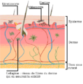
Peau modifie.png 453 × 455; 22 KB
-
Peau.png 332 × 329; 26 KB
-
Pell. 150X. Tricròmic de Masson. IMG 2511.JPG 453 × 619; 78 KB
-
Piel y cabello vs feolamina.jpg 300 × 275; 27 KB
-
Piel.png 498 × 493; 272 KB
-
Pl. 88 Bis. Anatomie Microscopique. Des Papilles De La Peau. Wellcome L0077001.jpg 4,876 × 7,056; 9.34 MB
-
Plate 88 Ter. Anatomie Microscopique De La Peau. Wellcome L0077000.jpg 4,856 × 7,048; 8.25 MB
-
Plate 88, Tome III. Microscopic structure of the skin. Wellcome L0077003.jpg 6,952 × 4,932; 10.22 MB
-
Scalp cross section (negro).jpg 3,740 × 3,740; 7.89 MB
-
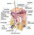
Skin anatomy esp.jpg 786 × 658; 290 KB
-
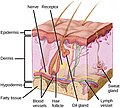
Skin anatomy.png 682 × 658; 283 KB
-
Skin dandruff viewed through a microscope.jpg 3,120 × 4,160; 879 KB
-
Skin Layers Unlabeled.jpg 3,500 × 3,500; 1.06 MB
-
Skin layers.png 1,282 × 997; 621 KB
-
Skin-hu.jpg 396 × 407; 98 KB
-
Skin.png 1,000 × 1,029; 658 KB
-
Skinlayers (español).png 438 × 385; 52 KB
-
Skinlayers (italiano).png 332 × 329; 42 KB
-
Skinlayers (italiano).svg 618 × 876; 281 KB
-
Skinlayers (labeled).svg 618 × 876; 241 KB
-
Skinlayers esp.jpg 683 × 876; 248 KB
-
Skinlayers pl.png 332 × 329; 111 KB
-
Skinlayers22.png 206 × 317; 25 KB
-
SLAUGA14-2. Ilona Varnelo. Oda. Iliustracijos.png 969 × 721; 1.11 MB
-
Structura pielii umane.png 1,486 × 1,529; 1.1 MB
-
The physiology and hygiene of the house in which we live (1887) (14801507653).jpg 1,496 × 1,078; 343 KB
-
The skin of mammals.jpg 550 × 700; 288 KB
-
Title page from T. Lewis, The blood vessels of the human skin Wellcome L0014159.jpg 1,162 × 1,680; 144 KB
-
Шкіра.png 396 × 407; 41 KB

