Category:Gray's Anatomy plates with help
Jump to navigation
Jump to search
Media in category "Gray's Anatomy plates with help"
The following 200 files are in this category, out of 330 total.
(previous page) (next page)-
 Abductor digiti minimi (foot).png 297 × 800; 159 KB
-
 Abductor hallucis.png 297 × 800; 164 KB
-
 Alaossissacri.PNG 500 × 333; 43 KB
-
 Angulusmandibulae.PNG 600 × 402; 23 KB
-
 Aqueductuscochleae.PNG 718 × 1,169; 147 KB
-
 Arcusanterioratlantis.png 500 × 235; 18 KB
-
 Arcusposterioratlantis.png 500 × 235; 18 KB
-
 Arcusvertebrae.png 450 × 287; 19 KB
-
 Asterion.PNG 759 × 599; 94 KB
-
 Auricularis anterior.png 428 × 410; 153 KB
-
 Auricularis posterior.png 428 × 410; 154 KB
-
 Auricularis superior.png 428 × 410; 156 KB
-
 Basion.PNG 718 × 1,169; 147 KB
-
 Brachialis.png 550 × 533; 236 KB
-
 Bregma.PNG 759 × 599; 94 KB
-
 Bulbospongiosus-Female.png 456 × 471; 171 KB
-
 Bulbospongiosus-Male.png 370 × 600; 246 KB
-
 Canalesalveolaresmaxillae.PNG 600 × 434; 46 KB
-
 Canaliculusmastoideus.PNG 550 × 495; 35 KB
-
 Canaliscaroticus.PNG 550 × 495; 35 KB
-
 Canaliscondylaris.PNG 550 × 529; 56 KB
-
 Canalisnervifacialis.PNG 550 × 338; 39 KB
-
 Capitulumhumeri.png 317 × 600; 111 KB
-
 Choana.PNG 718 × 1,169; 147 KB
-
 Chordaobliquamembranaeinterosseaeantebrachii.png 288 × 598; 258 KB
-
 Conchanasalismedius.PNG 600 × 501; 69 KB
-
 Coracobrachialis.png 550 × 533; 233 KB
-
 Cornucoccygeum.PNG 381 × 600; 39 KB
-
 Cornusacralis.PNG 381 × 600; 39 KB
-
 Corpusvertebrae.png 450 × 287; 19 KB
-
 Corrugator supercilii.png 428 × 410; 153 KB
-
 Coupe sagittale du genou (Gray 350).png 1,760 × 2,026; 828 KB
-
 Cristagalli.PNG 600 × 501; 70 KB
-
 Dacryon.PNG 650 × 466; 76 KB
-
 Depressor anguli oris.png 428 × 410; 154 KB
-
 Depressor labii inferioris.png 428 × 410; 154 KB
-
 Digastricus.png 536 × 568; 185 KB
-
 Elülsőkutacs.PNG 350 × 476; 48 KB
-
 Eminentiafrontalis.PNG 518 × 500; 39 KB
-
 Extensor digitorum longus.png 260 × 1,000; 146 KB
-
 Extensor hallucis longus.png 260 × 1,000; 141 KB
-
 Fissuraorbitalisinferior.PNG 582 × 600; 68 KB
-
 Flexor digitorum brevis.png 297 × 800; 163 KB
-
 Flexor-carpi-radialis.png 227 × 700; 97 KB
-
 Fonticulusmastoidalis.PNG 400 × 354; 40 KB
-
 Fonticulussphenoidalis.PNG 400 × 354; 40 KB
-
 Foramencaecumossisfrontalis.PNG 719 × 1,057; 149 KB
-
 Foramenjugulare.PNG 368 × 599; 268 KB
-
 Foramenparietalis.PNG 600 × 514; 65 KB
-
 Foramentransversarium.png 500 × 252; 17 KB
-
 Foramenvertebrale.png 450 × 287; 19 KB
-
 Foraminaintervertebralia.png 429 × 600; 49 KB
-
 Foraminasacraliaanteriora.PNG 525 × 550; 68 KB
-
 Foraminasacraliaposteriora.PNG 567 × 500; 45 KB
-
 Fossacanina.PNG 600 × 434; 46 KB
-
 Fossaglandulaelacrimalis.PNG 650 × 466; 76 KB
-
 Fossaolecrani.png 153 × 598; 77 KB
-
 Foveacostalisinferior.png 450 × 344; 21 KB
-
 Foveacostalissuperior.png 450 × 344; 20 KB
-
 Foveacostalistransversus-ar.png 450 × 344; 60 KB
-
 Foveacostalistransversus.png 450 × 344; 21 KB
-
 Frontoethmoidálisvarrat.PNG 408 × 600; 315 KB
-
 Frontálisvarrat.PNG 518 × 500; 66 KB
-
 Genioglossus.png 505 × 552; 135 KB
-
 Geniohyoideus.png 505 × 552; 131 KB
-
 Gnathion.PNG 500 × 463; 63 KB
-
 Gonion.PNG 500 × 463; 63 KB
-
 Gray1120-adrenal glands.png 683 × 900; 229 KB
-
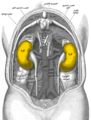 Gray1120-kidneys-ar.png 683 × 900; 546 KB
-
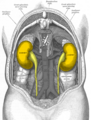
 Gray1120-kidneys.png 683 × 900; 251 KB
-
 Gray1120-ureters.png 683 × 900; 596 KB
-
 Gray1120-urinary-tract.png 683 × 900; 258 KB
-
 Gray164 - Zygomatic process of maxilla.png 400 × 379; 185 KB
-
 Gray188 - Temporal lines.png 500 × 463; 240 KB
-
 Gray193 - Cranial fossae-es.png 1,159 × 1,410; 1.6 MB
-
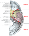 Gray193 - Cranial fossae.png 1,159 × 1,410; 1.48 MB
-
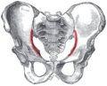 Gray241 - Arcuate line of ilium.png 500 × 404; 258 KB
-
 Gray378 (masseter highlight).png 423 × 403; 275 KB
-
 Gray415 - Adductor pollicis - red.png 327 × 353; 81 KB
-
 Gray415 - Flexor pollicis brevis - red.png 327 × 353; 79 KB
-
 Gray415 - Opponens digiti quinti - red.png 327 × 353; 79 KB
-
 Gray437-Musculus extensor digitorum brevis.png 258 × 325; 48 KB
-
 Gray437-Musculus extensor hallucis brevis.png 260 × 353; 50 KB
-
 Gray438-Musculus plantaris.png 280 × 900; 165 KB
-
 Gray439-Musculus flexor digitorum longus.png 222 × 900; 156 KB
-
 Gray439-Musculus popliteus.png 222 × 900; 162 KB
-
 Gray443-Musculus flexor digitorum brevis.png 297 × 800; 162 KB
-
 Gray533 es.png 800 × 630; 313 KB
-
 Gray702 anterior lobe of cerebellum.png 600 × 319; 313 KB
-
 Gray702 Cerebellar hemisphere.png 600 × 319; 350 KB
-
 Gray702 Cerebellar vermis.png 600 × 319; 308 KB
-
 Gray702 posterior lobe of cerebellum.png 600 × 319; 320 KB
-
 Gray702 primary fissure of cerebellum.png 600 × 319; 303 KB
-
 Gray703 Cerebellar vermis.png 600 × 350; 226 KB
-
 Gray812 pl.svg 371 × 800; 209 KB
-
 Gray812.svg 371 × 800; 45 KB
-
 Gray852 - Frontal process of maxilla.png 260 × 500; 117 KB
-
 Hanyintóalkariizom.PNG 337 × 1,000; 93 KB
-
 Hengeres borintóizom.PNG 194 × 598; 177 KB
-
 Hiatuscanalisfacialis.PNG 550 × 492; 52 KB
-
 Hosszú orsócsonti csuklófeszítő izom.PNG 196 × 599; 147 KB
-
 Hosszúhüvelykujj-hajlítóizom.PNG 337 × 1,000; 93 KB
-
 Hyoglossus.png 505 × 552; 141 KB
-
 Hátulsókutacs.PNG 350 × 476; 48 KB
-
 Hüvelykujjizompárnája.PNG 442 × 600; 313 KB
-
 Iliostalis.png 625 × 1,179; 377 KB
-
 Incisuraradialisulnae.png 215 × 600; 118 KB
-
 Incisuravertebralis.png 449 × 274; 27 KB
-
 Inferiortympanicuscanaliculus.PNG 550 × 495; 34 KB
-
 Inion.PNG 550 × 529; 92 KB
-
 Ischiocavernosus-female.png 455 × 470; 169 KB
-
 Ischiocavernosus-male.png 370 × 600; 249 KB
-
 Kisujjpárna.PNG 442 × 600; 315 KB
-
 Kisujjtávlítóizom(láb).PNG 222 × 598; 227 KB
-
 Laminaarcusvertebrae.png 450 × 287; 19 KB
-
 Levator anguli oris.png 428 × 410; 153 KB
-
 Levator ani.png 455 × 470; 172 KB
-
 Levator labii superioris.png 428 × 410; 158 KB
-
 Levator scapulae.png 733 × 1,156; 566 KB
-
 Levatores costarum.png 451 × 562; 218 KB
-
 Ligamentumcollateralecarpiradiale.PNG 509 × 466; 85 KB
-
 Ligamentumpisohamatum.png 509 × 466; 85 KB
-
 Ligamentumpisometacarpeum.png 509 × 466; 85 KB
-
 Ligamentumsacrococcygeumanterius.png 466 × 600; 476 KB
-
 Limites artère axillaire.png 1,361 × 1,196; 923 KB
-
 Longissimus.png 625 × 1,179; 379 KB
-
 Longus capitis.png 443 × 600; 148 KB
-
 Longus colli.png 443 × 600; 153 KB
-
 Lumbricales (hand).png 652 × 900; 292 KB
-
 Lumbricales pedis.png 330 × 800; 146 KB
-
 Massalateralisatlantis.png 500 × 235; 18 KB
-
 Mentalis.png 428 × 410; 153 KB
-
 Modiolus (face).PNG 531 × 600; 342 KB
-
 Multifidi.png 432 × 692; 263 KB
-
 Muscles rhomboïdes.jpg 733 × 1,298; 478 KB
-
 Musculus abductor digiti minimi (Hand).png 636 × 882; 131 KB
-
 Musculus abductor pollicis brevis - red.png 637 × 887; 319 KB
-
 Musculus abductor pollicis brevis.png 636 × 882; 279 KB
-
 Musculus adductor hallucis.png 361 × 800; 166 KB
-
 Musculus brachioradialis.PNG 194 × 598; 177 KB
-
 Musculus cremaster.png 564 × 550; 267 KB
-
 Musculus flexor digiti minimi brevis (foot).png 361 × 800; 154 KB
-
 Musculus flexor hallucis brevis.png 361 × 800; 159 KB
-
 Musculus flexor pollicis brevis - red.png 637 × 887; 338 KB
-
 Musculus frontalis.png 620 × 700; 308 KB
-
 Musculus occipitalis.png 620 × 700; 313 KB
-
 Musculus omohyoideus.png 600 × 477; 141 KB
-
 Musculus opponens pollicis - red.png 637 × 887; 347 KB
-
 Musculus psoas minor.png 354 × 580; 182 KB
-
 Musculus splenius capitis marked.png 733 × 1,156; 569 KB
-
 Musculusabductordigitiminimi(hand).png 434 × 599; 278 KB
-
 Musculusabductorpollicisbrevis.png 434 × 599; 277 KB
-
 Musculusabductorpollicislongus.png 234 × 598; 160 KB
-
 Musculusadductorpollicis.png 685 × 591; 133 KB
-
 Musculusanconeus.png 196 × 599; 136 KB
-
 Musculusantitragicus.png 400 × 548; 50 KB
-
 Musculusaryepiglotticus.png 404 × 600; 46 KB
-
 Musculusarytenoideus.png 363 × 500; 43 KB
-
 Musculusauricularisanterior.png 531 × 600; 386 KB
-
 Musculusauricularisposterior.png 531 × 600; 386 KB
-
 Musculusauricularissuperior.png 531 × 600; 387 KB
-
 Musculusbrachialis.png 550 × 533; 70 KB
-
 Musculusconstrictorpharyngisinferior.png 360 × 599; 278 KB
-
 Musculusconstrictorpharyngismedius.png 360 × 599; 276 KB
-
 Musculusconstrictorpharyngissuperior.png 360 × 599; 277 KB
-
 Musculuscorrugatorsupercilii.png 535 × 450; 83 KB
-
 Musculuscricoarytenoideuslateralis.png 404 × 600; 46 KB
-
 Musculuscricoarytenoideusposterior.png 404 × 600; 46 KB
-
 Musculuscricopharyngeus.PNG 400 × 665; 85 KB
-
 Musculuscricothyreoideus.png 404 × 600; 46 KB
-
 Musculusdepressorangulioris.png 619 × 391; 711 KB
-
 Musculusdepressorlabiiinferioris.png 619 × 391; 711 KB
-
 Musculusdepressorseptinasi.png 619 × 391; 711 KB
-
 Musculusdilatatornarisanterior.png 619 × 391; 711 KB
-
 Musculusdilatatornarisposterior.png 619 × 391; 711 KB
-
 Musculusdilatatornarisposterior2.PNG 620 × 700; 85 KB
-
 Musculusextensordigitiminimi.png 196 × 599; 136 KB
-
 Musculusextensorindicisproprius.png 234 × 598; 159 KB
-
 Musculusextensorpollicisbrevis.png 234 × 598; 159 KB
-
 Musculusextensorpollicislongus.png 234 × 598; 159 KB
-
 Musculusflexorcarpiulnaris.png 194 × 598; 179 KB
-
 Musculusflexordigitiminimibrevismanus.png 434 × 599; 277 KB
-
 Musculusflexordigitorumsuperficialis.png 194 × 598; 177 KB
-
 Musculusflexorpollicisbrevis.png 434 × 599; 275 KB
-
 Musculusfrontalis.png 619 × 391; 711 KB
-
 Musculusgenioglossus.png 594 × 600; 307 KB
-
 Musculushelicismajor.png 400 × 548; 50 KB
-
 Musculushelicisminor.png 400 × 548; 50 KB
-
 Musculushyoglossus.png 594 × 600; 305 KB
-
 Musculuslevatorangulioris.png 531 × 600; 383 KB
-
 Musculuslevatorlabiisuperioris.png 531 × 600; 385 KB
-
 Musculuslevatorlabiisuperiorisalaequenasi.png 531 × 600; 388 KB
-
 Musculuslevatorvelipalatini.png 500 × 600; 323 KB
-
 Musculuslongitudinalisinferiorlinguae.png 594 × 600; 307 KB
-
 Musculuslongitudinalissuperiorlinguae.png 500 × 300; 53 KB
-
 Musculusmasseter.png 531 × 600; 386 KB
-
 Musculusmentalis.png 531 × 600; 388 KB
-
 Musculusnasalis.png 531 × 600; 388 KB
-
 Musculusoccipitalis.png 531 × 600; 388 KB
-
 Musculusopponensdigitiminimi.png 202 × 599; 128 KB


.png)


.png)


.png)


.PNG)


.png)


.png)


.png)


.png)
.PNG)