Category:Gram positive
Jump to navigation
Jump to search
Subcategories
This category has the following 2 subcategories, out of 2 total.
G
S
- Sarcina (bacterium) (10 F)
Media in category "Gram positive"
The following 54 files are in this category, out of 54 total.
-
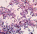
1. Грамположительные палочки.jpg 2,801 × 2,425; 1.76 MB
-
2. Грамположительные палочки.jpg 2,985 × 2,745; 1.92 MB
-
-
-
A-Trigger-Enzyme-in-Mycoplasma-pneumoniae-Impact-of-the-Glycerophosphodiesterase-GlpQ-on-Virulence-ppat.1002263.s010.ogv 6.1 s, 1,920 × 1,080; 2.65 MB
-
-
Bacillus cereus (15469201572).jpg 1,756 × 988; 2.47 MB
-
Blood culture gram stain of Gram positive cocci.jpg 1,213 × 1,148; 67 KB
-
Candida in Gram staining.jpg 3,264 × 2,448; 1.07 MB
-
Chronic-Alcohol-Exposure-Renders-Epithelial-Cells-Vulnerable-to-Bacterial-Infection-pone.0054646.s004.ogv 11 s, 1,404 × 590; 15.41 MB
-
Chronic-Alcohol-Exposure-Renders-Epithelial-Cells-Vulnerable-to-Bacterial-Infection-pone.0054646.s005.ogv 6.7 s, 1,404 × 590; 11.32 MB
-
Chronic-Alcohol-Exposure-Renders-Epithelial-Cells-Vulnerable-to-Bacterial-Infection-pone.0054646.s006.ogv 6.8 s, 1,404 × 606; 7.59 MB
-
Chronic-Alcohol-Exposure-Renders-Epithelial-Cells-Vulnerable-to-Bacterial-Infection-pone.0054646.s007.ogv 4.1 s, 1,404 × 592; 6.63 MB
-
Chronic-Alcohol-Exposure-Renders-Epithelial-Cells-Vulnerable-to-Bacterial-Infection-pone.0054646.s008.ogv 6.7 s, 1,404 × 590; 8.86 MB
-
Chronic-Alcohol-Exposure-Renders-Epithelial-Cells-Vulnerable-to-Bacterial-Infection-pone.0054646.s009.ogv 8.1 s, 1,404 × 590; 19.07 MB
-
Chronic-Alcohol-Exposure-Renders-Epithelial-Cells-Vulnerable-to-Bacterial-Infection-pone.0054646.s010.ogv 4.1 s, 1,404 × 590; 7.22 MB
-
Chronic-Alcohol-Exposure-Renders-Epithelial-Cells-Vulnerable-to-Bacterial-Infection-pone.0054646.s011.ogv 12 s, 1,404 × 412; 10.59 MB
-
Chronic-Alcohol-Exposure-Renders-Epithelial-Cells-Vulnerable-to-Bacterial-Infection-pone.0054646.s012.ogv 12 s, 1,404 × 412; 6.99 MB
-
Coloração de Gram.png 452 × 452; 61 KB
-
Deinococcus geothermalis cells.jpg 640 × 531; 122 KB
-
Gram -Positive and Negative Bacteria.jpg 4,000 × 2,250; 1.16 MB
-
Gram Negative Rods in Gram Stained Smear of Culture.jpg 4,000 × 2,250; 1.14 MB
-
Gram positive bacilli.jpg 2,068 × 1,336; 1.48 MB
-
Gram positive cocci in chains.jpg 4,000 × 2,250; 2.09 MB
-
Gram positive cocci in clusters.jpg 1,128 × 726; 702 KB
-
Gram Positive Cocci in singles, pairs and clusters.jpg 4,160 × 2,340; 2.2 MB
-
Gram positive cocci.jpg 1,388 × 868; 509 KB
-
Gram positive diagram.pdf 3,375 × 2,297; 194 KB
-
Gram Positive.png 445 × 382; 127 KB
-
Gram-positive bacteria and pus cells.jpg 1,758 × 1,416; 780 KB
-
GramPositiveStain.jpg 1,787 × 1,582; 866 KB
-
Labori algoritm, et eristada grampositiivseid baktereid.png 663 × 445; 37 KB
-
Leuconostoc mesenteroides Gram Staining.jpg 4,000 × 3,000; 1.32 MB
-
LexA gram positive bacteria sequence logo.png 680 × 188; 11 KB
-
Lysozyme agar diffusion assay.png 4,770 × 2,275; 18.94 MB
-
-
-
-
Pus in Gram stain showing Gram positive cocci in singles, pairs and clusters.jpg 4,000 × 2,250; 1.44 MB
-
-
-
-
Revestimentos B.png 291 × 452; 52 KB
-
Rhodococcus spp..jpg 1,600 × 1,200; 672 KB
-
S. agalactiae in Gram staining of old culture.jpg 4,000 × 2,250; 1.06 MB
-
Staphylococcus haemolyticus in Gram staining.jpg 4,000 × 2,250; 1.43 MB
-
Staphylococcus hominis colony morphology on blood agar.jpg 4,000 × 3,000; 1.3 MB
-
Staphylococcus hominis Gram staining.jpg 4,000 × 2,250; 1.53 MB
-
Streptococcus agalactiae Gram staining of culture.jpg 4,000 × 2,250; 958 KB
-
Variety of Gram positive and Negative bacteria in Gram stain of Sewage.jpg 4,000 × 2,250; 1.61 MB
-
Yeast cells in Gram staining of Candida growth on SDA.jpg 4,160 × 2,340; 1.46 MB
-
Yeast cells of Candida albicans in Gram staining of culture microscopy.jpg 4,160 × 2,340; 2.15 MB


