Category:Gonads
Jump to navigation
Jump to search
endocrine gland that produces the gametes of an organism | |||||
| Upload media | |||||
| Instance of |
| ||||
|---|---|---|---|---|---|
| Subclass of |
| ||||
| |||||
Subcategories
This category has the following 8 subcategories, out of 8 total.
Media in category "Gonads"
The following 88 files are in this category, out of 88 total.
-
A treatise on zoology (1900) (14760329746).jpg 1,124 × 2,582; 304 KB
-
-
-
Amphioxus pharyngeal Basket and gonads notochord nerve chord.jpg 1,280 × 720; 67 KB
-
Amphioxus XS showing pharynx and gonads.jpg 1,280 × 720; 78 KB
-
Amphioxus xs through pharyngeal basket and gonads.jpg 1,280 × 720; 79 KB
-
Archimollusc-num.svg 640 × 420; 8.98 MB
-
Astacopsidrilus hibernicus (10.11646-zoosymposia.17.1.6) Figure 3.png 3,908 × 2,080; 825 KB
-
Aurelia aurita 2.jpg 2,845 × 2,845; 1.59 MB
-
Aurelia Aurita.JPG 3,968 × 2,976; 2.85 MB
-
Cartwright et al. 2007 f8.png 3,150 × 1,296; 5.68 MB
-
Celula gonadotropa FSH.jpg 760 × 614; 233 KB
-
Chicken PGCs.png 2,708 × 1,259; 737 KB
-
-
-
Clytia hemisphaerica female gonad.svg 512 × 285; 10 KB
-
Development of primordial germ cells (PGCs) and expression of markers. Chicken.png 2,756 × 1,668; 442 KB
-
Differentiation sexuel.png 1,297 × 2,444; 489 KB
-
Ectopic Expression of Male Factors, SOX9 and FGF9, in XX Wnt4 minus-minus Gonads.png 2,012 × 2,072; 4.42 MB
-
Epistatic Relationship of Sry, Fgf9, and Sox9.png 2,398 × 3,355; 2.7 MB
-
Female gonads from rainbow trout (Oncorhynchus mykiss).jpg 1,080 × 810; 131 KB
-
GermCellMigration.jpg 726 × 660; 50 KB
-
Gonad Precursor Migration.png 1,668 × 1,668; 474 KB
-
Gonadi Paracentrotus lividus riccio di mare adventurediving.it.jpg 1,000 × 750; 203 KB
-
Gonadotrofas Capilar.png 1,016 × 1,026; 1.7 MB
-
Gonadotrofas FSH LH.png 686 × 336; 274 KB
-
Gonadotropas Capilar.png 584 × 602; 316 KB
-
Gonadotropas Stellates.jpg 776 × 628; 245 KB
-
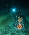
Ice planet and antarctic jellyfish (cropped).jpg 1,742 × 2,066; 2.49 MB
-
Image from page 337 of "Chordate morphology" (1962) (19989513674).jpg 1,870 × 794; 344 KB
-
Interdependent Relationship between Fgf9 and Sox9.png 3,128 × 3,477; 3.41 MB
-
Internal structures of Concavicaris submarinus (PIMUZ 37349).jpg 1,350 × 926; 142 KB
-
Internal urinogenital organs of free-martin.jpg 986 × 1,572; 676 KB
-
Internal urinogenital organs of male twin.jpg 998 × 1,642; 765 KB
-
Lancetnikinside.png 1,600 × 529; 243 KB
-
Leucochloridium paradoxum metacercaria from Heckert 1889 plate1 fig5.png 541 × 795; 488 KB
-
-
Male and female gonads 1.png 2,220 × 1,152; 400 KB
-
Mawia benovici DSC1588.jpg 1,920 × 1,280; 1.45 MB
-
Medusae of the world (1910) (14779678514).jpg 1,296 × 844; 178 KB
-
Medusae of world-vol03 fig332a Craterlophus tethys.jpg 724 × 578; 158 KB
-
Melicertissa antrichardsoni (10.3897-zookeys.783.26862) Figure 3.jpg 1,217 × 1,740; 787 KB
-
Montgomery Plethodon cinereus larva.jpg 1,383 × 2,133; 586 KB
-
Moon Jellies (Aurelia aurita) (7153396279).jpg 4,917 × 3,278; 12.84 MB
-
Moon jellyfish - geograph.org.uk - 851268.jpg 640 × 480; 163 KB
-
Moon jellyfish with six gonads.jpg 2,668 × 2,669; 3.63 MB
-
Mutual Antagonism between Fgf9 and Wnt4.png 1,788 × 2,128; 2.41 MB
-
Nausithoe maculata (10.3897-zookeys.984.56380) Figure 6.jpg 1,512 × 675; 457 KB
-
Nematode C. elegans Anatomy Relating to the Control of Meiotic Maturation.jpg 1,800 × 2,281; 2.41 MB
-
Nematode C. elegans gonadogenesis.jpg 1,792 × 1,193; 557 KB
-
Opposing Signals Regulate Sex Determination in the Bipotential Gonad.png 2,624 × 3,989; 634 KB
-
Ovaries of Cyprinus carpio.png 1,348 × 1,290; 3 MB
-
Pantelozetes cavaticus (10.3897-subtbiol.16.8609) Figure 3.jpg 1,182 × 886; 936 KB
-
Paralovenia yongalensis (10.3897-zookeys.783.26862) Figure 5.jpg 1,170 × 1,794; 887 KB
-
-
-
-
-
Puberty-Hormonal control.jpg 2,880 × 2,080; 1.53 MB
-
Quain's elements of anatomy (1882) (14761555234).jpg 1,364 × 538; 107 KB
-
Sertoli Cell Precursors Switch from Expression of Male to Female Pathway Genes.png 2,984 × 2,476; 5.45 MB
-
Sex-differentiation-Image004.jpg 800 × 674; 114 KB
-
Stage- and Cell-Specific Expression of FGF9 in Embryonic Gonads.png 2,409 × 2,591; 3.77 MB
-
Supporting-like PAX8+ (sPAX8) gonadal lineage forms the rete testis.jpg 1,081 × 1,313; 1.45 MB
-
Tainha Fish eggs.jpg 3,200 × 2,551; 4.15 MB
-
Tellinidae (10.3897-zookeys.705.12888) Figure 4.jpg 1,200 × 1,182; 560 KB
-
The Biological bulletin (20187623059).jpg 1,720 × 2,046; 450 KB
-
The Journal of experimental zoology (1918) (14594487998).jpg 1,920 × 2,732; 995 KB
-
The uterus differentiates from the fetal Müllerian ducts..jpg 494 × 477; 76 KB
-
Tissue-resident macrophages in the developing testes.jpg 2,119 × 1,372; 2.4 MB
-
Transcriptional, spatiotemporal and paracrine signatures of human pregranulosa cells.jpg 2,118 × 1,595; 1.91 MB
-
पौगंडावस्थेतील संप्रेरक-नियंत्रण - Puberty-Hormonal control.jpg 2,880 × 2,080; 497 KB


