Category:Golgi apparatus
Jump to navigation
Jump to search
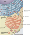
cell organelle that packages proteins for export  Esquema del proceso secretor desde el retículo endoplasmático (naranja) al aparato de Golgi (rosa). 1. Membrana nuclear. 2. Poro nuclear. 3. Retículo endoplasmático rugoso (RER). 4. 4. Retículo endoplasmático liso (REL). 5. Ribosoma unido al RER. 6. Macromoléculas. 7. Vesícula de transporte. 8. Aparato de Golgi. 9. Cara de la cisterna del aparato de Golgi. 10. Cara de transporte del aparato de Golgi. 11. Cisterna de lípidos. | |||||
| Upload media | |||||
| Instance of |
| ||||
|---|---|---|---|---|---|
| Subclass of |
| ||||
| Part of | |||||
| Named after | |||||
| Discoverer or inventor | |||||
| Has part(s) |
| ||||
| |||||
Subcategories
This category has the following 3 subcategories, out of 3 total.
Media in category "Golgi apparatus"
The following 62 files are in this category, out of 62 total.
-
0314 Golgi Apparatus a ar.png 579 × 708; 357 KB
-
0314 Golgi Apparatus a en.png 579 × 708; 368 KB
-
0314 Golgi Apparatus a ru.png 605 × 708; 436 KB
-
0314 Golgi Apparatus b en.png 521 × 435; 234 KB
-
0314 Golgi Apparatus.jpg 1,124 × 754; 494 KB
-
201601 golgi body.png 659 × 434; 134 KB
-
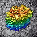
Algal Golgi body, 3D reconstruction (30466939665).jpg 2,043 × 2,043; 478 KB
-
Aparato-Golgi.jpg 1,355 × 773; 180 KB
-
ApicoBasoc.jpg 331 × 530; 58 KB
-
Base V2-01.jpg 1,754 × 839; 306 KB
-
Blausen 0435 GolgiApparatus.png 1,600 × 1,296; 927 KB
-
Cellorganeller pic swe 27-05-2007.png 603 × 481; 100 KB
-
Classificació SNAREs wiki.png 2,048 × 1,536; 313 KB
-
Coated VacV forming.jpg 1,654 × 1,649; 1.13 MB
-
Drawing of Golgi Apparatus.jpg 2,048 × 1,324; 867 KB
-
Endomembrane system ar.png 612 × 486; 91 KB
-
Esocitosi.gif 829 × 560; 284 KB
-
Esquema distribucio.png 887 × 479; 149 KB
-
Estructura STX-6.png 1,798 × 970; 54 KB
-
Flipases flopases i escramblases.png 885 × 473; 149 KB
-
Flipases.png 908 × 546; 156 KB
-
Glycan processing in the ER and Golgi.png 4,438 × 2,830; 300 KB
-
Golgi 3D Ostreococcus.png 2,004 × 529; 1.17 MB
-
Golgi Apparatus.jpg 891 × 602; 172 KB
-
Golgi bifurcado Epididimo.png 2,054 × 1,018; 2.23 MB
-
Golgi body.JPG 487 × 181; 22 KB
-
Golgi Cinta Complejo.png 903 × 906; 767 KB
-
Golgi Cisterna Membrana.png 616 × 618; 438 KB
-
Golgi Cisterna.png 586 × 586; 354 KB
-
Golgi disposicion Glia-radial Rata.jpg 556 × 1,788; 288 KB
-
Golgi in the cytoplasm of a macrophage in the alveolus (lung) - TEM.jpg 640 × 480; 161 KB
-
Golgi localización.png 1,224 × 349; 431 KB
-
Golgi Pila.png 1,366 × 760; 1.08 MB
-
Golgi portadores.jpg 1,929 × 1,335; 591 KB
-
Golgi ribbon humana.png 1,161 × 609; 676 KB
-
Golgi secretions.png 432 × 301; 68 KB
-
Golgi ubicacion Glia-radial Rata.jpg 700 × 1,788; 381 KB
-
Golgi ubicación.png 1,920 × 485; 502 KB
-
Golgi.JPG 433 × 269; 17 KB
-
Golgi.png 1,054 × 516; 426 KB
-
GolgiAFMc.jpg 407 × 456; 65 KB
-
GolgiFboxc.jpg 530 × 288; 74 KB
-
Golgijev stanica.png 200 × 200; 39 KB
-
GolgilTGNc.jpg 649 × 465; 118 KB
-
GolgiMovieArrorleftc.jpg 336 × 167; 37 KB
-
GolgiMovieArrowc.jpg 490 × 155; 47 KB
-
GolgiStrucGenec.jpg 506 × 271; 74 KB
-
GolgiTGNcolorc.jpg 532 × 297; 81 KB
-
HeLa-I.jpg 2,400 × 1,999; 2.15 MB
-
Human leukocyte, showing golgi - TEM.jpg 640 × 480; 97 KB
-
Local de brotamento de vesícula revestida de clatrina.jpg 2,048 × 1,536; 215 KB
-
Meaning-golgi-apparatus-800x800.jpg 877 × 500; 51 KB
-
Model for Golgi Formation.jpg 497 × 339; 43 KB
-
N-linked protein glycosylation in the ER.svg 2,662 × 1,018; 164 KB
-
Nucleus ER golgi ex.jpg 534 × 426; 49 KB
-
Nucleus ER golgi.jpg 492 × 565; 62 KB
-
Protein STX6 PDB 1lvf.png 895 × 527; 215 KB
-
Rol de les flipases en la biogènesi vesicular.jpg 430 × 201; 26 KB
-
Sekretorischer weg.jpg 604 × 187; 26 KB
-
The Biological bulletin (19755382264).jpg 1,958 × 1,324; 651 KB
-
Апарат Гольджи в нервных клетках спинального ганглия котенка.jpg 1,074 × 1,080; 160 KB


