Category:Frontal sinuses
Jump to navigation
Jump to search
one of the four pairs of paranasal sinuses that are situated behind the brow ridges | |||||
| Upload media | |||||
| Instance of |
| ||||
|---|---|---|---|---|---|
| Subclass of |
| ||||
| Part of | |||||
| |||||
Media in category "Frontal sinuses"
The following 47 files are in this category, out of 47 total.
-
3D Cinematic Rendering reconstructions of the depressed frontal fracture.png 1,058 × 782; 800 KB
-
3D Cinematic Rendering reconstructions of the depressed frontal fracture2.png 1,058 × 716; 583 KB
-
Crane4 Foramen magnum.png 2,431 × 2,761; 6.83 MB
-
Crane4.png 2,431 × 2,761; 6.96 MB
-
Cranium - sinus frontalis detail.jpg 3,616 × 3,000; 3.08 MB
-
Einseitige Aplasie der Stirnhoehle 44W - CT axial - 001.jpg 1,599 × 652; 224 KB
-
Einseitige Hypo- Aplasie Stirnhoehle 80M - CT axial und coronar - 001.jpg 1,546 × 683; 153 KB
-
Fraktur Stirnhoehle axial.png 848 × 772; 345 KB
-
Frontal bone sinuses.jpg 300 × 330; 65 KB
-
Frontal sinus inflamation.jpg 1,047 × 1,276; 677 KB
-
Frontal sinus.jpg 960 × 720; 75 KB
-
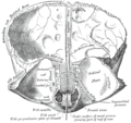
Gray1199.png 322 × 500; 33 KB
-
Gray135.png 550 × 518; 64 KB
-
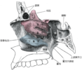
Gray153 zh.png 600 × 501; 200 KB
-
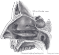
Gray153.png 600 × 501; 62 KB
-
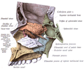
Gray159.png 700 × 503; 78 KB
-
Gray194.png 650 × 420; 84 KB
-
Gray195.png 500 × 433; 27 KB
-
Gray196.png 600 × 428; 43 KB
-
Gray855 zh.png 500 × 468; 185 KB
-
Gray855.png 500 × 468; 53 KB
-
Gray994 zh.png 600 × 861; 329 KB
-
Gray994.png 600 × 861; 112 KB
-
He cranium sectioned. Verso- The skull sectioned 1489.jpg 349 × 480; 62 KB
-
Illu nose nasal cavities.png 1,456 × 844; 337 KB
-
Illu09 sinuses.jpg 478 × 264; 41 KB
-
Lateraler Auslaeufer des Sinus frontalis 47W - CR ap - 001.jpg 1,072 × 1,124; 114 KB
-
Lateraler Auslaeufer des Sinus frontalis 47W - CR seitlich - 001.jpg 1,367 × 1,124; 129 KB
-
Leonardo Skull.jpg 360 × 426; 76 KB
-
Nasennebenhöhlen.gif 600 × 428; 66 KB
-
NNH im Roentgen frontal Annotation.png 678 × 962; 440 KB
-
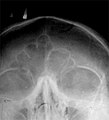
NNH im Roentgen frontal.png 678 × 962; 393 KB
-
Occipital bone.jpg 960 × 720; 76 KB
-
Orbite et Sinus maxillaire.png 735 × 608; 268 KB
-
Pferdeschädel.jpg 1,006 × 325; 47 KB
-
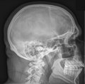
Schaedel im Röntgen seitlich.png 1,092 × 1,078; 640 KB
-
Sinus frontalis in red.png 690 × 592; 62 KB
-
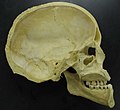
Skull - midsaggital section P.2005.jpg 1,024 × 937; 62 KB
-
Skull inner surface.jpg 2,409 × 3,134; 5.27 MB
-
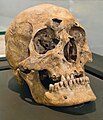
Skull of Odwinus.jpg 1,329 × 1,546; 1.39 MB
-
Slide3rr.JPG 960 × 720; 118 KB
-
Sobo 1909 101.png 2,068 × 1,588; 9.41 MB
-
Sobo 1909 102.png 1,928 × 1,628; 9 MB
-
Some points in the surgery of the brain and its membranes (1907) (14586124588).jpg 2,064 × 1,330; 580 KB
-
Some points in the surgery of the brain and its membranes (1907) (14586125668).jpg 2,096 × 1,482; 624 KB
-
Teviec profil droit.jpg 3,048 × 3,056; 5.78 MB
-
View of a Skull III.jpg 813 × 992; 486 KB


