Category:Files from Berkshire Community College Bioscience Image Library Flickr stream uploaded by Netha Hussain
Jump to navigation
Jump to search
Media in category "Files from Berkshire Community College Bioscience Image Library Flickr stream uploaded by Netha Hussain"
The following 164 files are in this category, out of 164 total.
-
 About the Bioscience Image Library (37088502443).jpg 3,264 × 2,448; 2.8 MB
-
 Ameboid Protozoa Amoeba proteus (37175354480).jpg 3,264 × 1,840; 744 KB
-
 Amoeboid Protozoa Cytoplasmic Streaming in Amoeba proteus (34306696281).jpg 1,920 × 1,080; 114 KB
-
 Aquatic Monocot Root Elodea (35171859074).jpg 3,264 × 1,840; 1.23 MB
-
 Aquatic Monocot Root Stele in Elodea (36011419685).jpg 3,264 × 1,840; 1.29 MB
-
 Aquatic Monocot Stem Aerenchyma in Potamogeton (35576919670).jpg 3,264 × 1,840; 1.5 MB
-
 Aquatic Monocot Stem Aerenchyma in Potamogeton (35576920640).jpg 3,264 × 1,840; 1.26 MB
-
 Aquatic Monocot Stem Central Stele in Elodea (37626081871).jpg 3,264 × 1,840; 1.04 MB
-
 Aquatic Monocot Stem Cortex in Elodea (36915455834).jpg 3,264 × 1,840; 1.26 MB
-
 Aquatic Monocot Stem Dermal Tissues in Elodea (37626081061).jpg 3,264 × 1,840; 1.04 MB
-
 Aquatic Monocot Stem Elodea (35570465200).jpg 3,264 × 1,840; 4.68 MB
-
 Aquatic Monocot Stem Elodea (35570465720).jpg 3,264 × 1,840; 5.95 MB
-
 Aquatic Monocot Stem Epidermis in Elodea (37626079451).jpg 3,264 × 1,840; 3.48 MB
-
 Aquatic Monocot Stem Epidermis in Potamogeton (35576920200).jpg 3,264 × 1,840; 1.19 MB
-
 Aquatic Monocot Stem Potamogeton (35576923160).jpg 3,264 × 1,840; 6.61 MB
-
 Aquatic Monocot Stem Potamogeton (35576924860).jpg 3,264 × 1,840; 1.66 MB
-
 Aquatic Monocot Stem Stele in Elodea (36915456804).jpg 3,264 × 1,840; 1,012 KB
-
 Aquatic Monocot Stem Stele in Elodea (36915457514).jpg 3,264 × 1,840; 1.15 MB
-
 Aquatic Monocot Stem Stele in Potamogeton (35576925980).jpg 3,264 × 1,840; 2.21 MB
-
 Aquatic Monocot Stem Vascular Bundles in Potamogeton (35576921420).jpg 3,264 × 1,840; 1.76 MB
-
 Border Between Horny and Floury Endosperm in Zea Mays Embryo (46915319864).jpg 3,264 × 1,840; 1.01 MB
-
 Bran and Aleurone Layer in Zea Mays Embryo by Phase Contrast (40724991623).jpg 3,264 × 1,840; 994 KB
-
-

-
 Ciliated Protozoa Colpidium colpoda (33627323753).jpg 1,920 × 1,080; 249 KB
-
 Ciliated Protozoa Vorticella (43855973655).jpg 1,920 × 1,080; 78 KB
-
 Ciliated Protozoa Vorticella (44046567434).jpg 3,264 × 1,840; 489 KB
-
 Ciliated Protozoa Vorticella (44046569784).jpg 1,920 × 1,080; 88 KB
-
 Ciliated Protozoa Vorticella (44716054772).png 1,096 × 1,084; 952 KB
-
 Ciliated Protozoa Vorticella (44716055852).jpg 1,920 × 1,080; 77 KB
-
 Closed Collateral Vascular Bundle in Monocot Stem (35995829090).jpg 3,264 × 1,840; 5.5 MB
-
 Coleoptile tip and Plumule in Zea Mays Embryo (46915320474).jpg 3,264 × 1,840; 1.25 MB
-
 Cortex Parenchyma in Herbaceous Dicot Ranunculus (34758991731).jpg 3,264 × 1,840; 1.2 MB
-
 Cotyledon, Coleoptile and Plumule in Zea Mays Embryo (47638340702).jpg 3,264 × 1,840; 1.35 MB
-
 Digestive and Absorptive Tissues of the Cotyledon (47691344831).jpg 3,264 × 1,840; 5.76 MB
-
 Endosperm and Cotyledon in Zea Mays Embryo (40724990373).jpg 3,264 × 1,840; 1.42 MB
-
 Endosperm, Protective Cotelydon Tip and Bran in Zea Mays Embryo (46915320104).jpg 3,264 × 1,840; 1.07 MB
-
 Epithelial Tissues Simple Columnar Epithelium (41006485014).jpg 3,264 × 1,840; 6.55 MB
-
 Epithelial Tissues Simple Squamous Epithelium (frog) (27847646938).jpg 3,264 × 1,840; 4.75 MB
-
 Epithelial Tissues Simple Squamous Epithelium (frog) (27847648428).jpg 3,264 × 1,840; 7.4 MB
-
 Epithelial Tissues Simple Squamous Epithelium (frog) (39909208420).jpg 3,264 × 1,840; 5.82 MB
-
 Flagellated Protozoa Radiolaria (34779750100).jpg 3,264 × 1,840; 553 KB
-
 Gymnosperm Leaves Vascular Bundle in Five Needle Pinus (36325130400).jpg 3,264 × 1,837; 4.52 MB
-
 Herbaceous Dicot Root Closed Vascular Bundle in Mature Ranunculus (35613584780).jpg 3,264 × 1,840; 1.25 MB
-
 Herbaceous Dicot Root Mature Ranunculus (35613584240).jpg 3,264 × 1,840; 2.04 MB
-
 Herbaceous Dicot Root Mature Ranunculus (35613585260).jpg 3,264 × 1,840; 1.13 MB
-
 Herbaceous Dicot root Ranunculus (34727536412).jpg 3,264 × 1,840; 5.59 MB
-
 Herbaceous Dicot Root Ranunculus (34850808736).jpg 3,264 × 1,840; 5.01 MB
-
 Herbaceous Dicot Stem Cucurbita (35207517850).jpg 3,264 × 1,840; 1.6 MB
-
 Herbaceous Dicot Stem Cucurbita (35490271331).jpg 3,264 × 1,840; 1.47 MB
-
 Herbaceous Dicot Stem Vascular Bundles in Cucurbita (35207532680).jpg 3,264 × 1,840; 1.15 MB
-
 Herbaceous Dicot Stem Xylem Vessels Cucurbita (35463815631).jpg 3,264 × 1,840; 1.17 MB
-
 Horny and Floury Endosperm in Zea Mays Embryo (47690779451).jpg 3,264 × 1,840; 2.06 MB
-
 Horny and Floury Endosperm in Zea Mays Embryo by Phase Contrast (40725881773).jpg 3,264 × 1,840; 1.61 MB
-
 Horny and Floury Endosperm in Zea Mays Embryo by Phase Contrast (46915319554).jpg 3,264 × 1,840; 2.31 MB
-
 Horny and Floury Endosperm of Zea Mays Embryo by Phase Contrast (47691345291).jpg 3,264 × 1,840; 1.87 MB
-
 Horny Endosperm in Zea Mays Embryo by Phase Contrast (40725880953).jpg 3,264 × 1,840; 1.84 MB
-
 IMG00539 seed and ovary coat (47636155012).jpg 3,264 × 1,840; 954 KB
-
 IMG00556 endosperm (47636154692).jpg 3,264 × 1,840; 2.04 MB
-
 IMG00557 endosperm (46773066895).jpg 3,264 × 1,840; 1.87 MB
-
 IMG00559 endosperm phase (33811606368).jpg 3,264 × 1,840; 1.84 MB
-
 IMG00570 cotyledon (46773065975).jpg 3,264 × 1,840; 1.3 MB
-
 IMG00572 pallisade mesophyll prevasc system (46773065445).jpg 3,264 × 1,840; 1.43 MB
-
 IMG00574 prtoective colyeoptile (46773064845).jpg 3,264 × 1,840; 1.16 MB
-
 IMG00581 hypocotyl regio (47636155602).jpg 3,264 × 1,840; 1.38 MB
-
 IMG00582 hypocotyle region (46773063875).jpg 3,264 × 1,840; 1.51 MB
-
 Junction Node at Hypotocotyl and Radicle in Zea Mays Embryo (33812368978).jpg 3,264 × 1,840; 1.61 MB
-
 Junction Node Between Hypotocotyl and Radicle in Zea Mays Embryo (47638340582).jpg 3,264 × 1,840; 5.65 MB
-
 Junction of Cotyledon, Hypocotyl and Plumule in Zea Mays Embryo (40724989523).jpg 3,264 × 1,840; 1.43 MB
-
 Junction of Hypocotyl and Plumule in Zea Mays Embryo (47636871162).jpg 3,264 × 1,840; 5.12 MB
-
 Lateral Root Origin (36223436805).jpg 3,264 × 1,840; 586 KB
-
 Lateral Root Origin (36223438925).jpg 3,264 × 1,840; 820 KB
-
 Lateral Root Origin (36223441325).jpg 3,264 × 1,840; 889 KB
-
 Lateral Root Origin (36223443845).jpg 3,264 × 1,840; 853 KB
-
 Monocot Epidermis in Smilax (35963298766).jpg 3,264 × 1,840; 1.12 MB
-
 Monocot Heavily Suberized Endodermis in Smilax (36004774435).jpg 3,264 × 1,840; 1.31 MB
-
 Monocot root Acorus (35141257384).jpg 3,264 × 1,840; 2.02 MB
-
 Monocot Root Acorus (35399384203).jpg 3,264 × 1,840; 6.54 MB
-
 Monocot Root Acorus (35592057590).jpg 3,264 × 1,840; 1.18 MB
-
 Monocot Root Acorus (35939628296).jpg 3,264 × 1,840; 1.4 MB
-
 Monocot Root Acorus (35939630426).jpg 3,264 × 1,840; 2.98 MB
-
 Monocot Root Acorus (36036866902).jpg 3,264 × 1,840; 4.33 MB
-
 Monocot Root Aerenchyma in Acorus (35399382813).jpg 3,264 × 1,840; 2.05 MB
-
 Monocot Root Casparian Strip in Acorus Vascular Bundle (35939627386).jpg 3,264 × 1,840; 1.45 MB
-
 Monocot Root Endodermis in Smilax (35615771550).jpg 3,264 × 1,840; 1.93 MB
-
 Monocot Root Epidermal Root Hair in Lilium (36045152715).jpg 3,264 × 1,840; 764 KB
-
 Monocot Root Epidermis and Cortex in Acorus (35399384763).jpg 3,264 × 1,840; 876 KB
-
 Monocot Root Epidermis and Cortex in Smilax (36004773605).jpg 3,264 × 1,840; 1.77 MB
-
 Monocot Root Epidermis and Cortex in Zea Prop Root (36008663576).jpg 3,264 × 1,840; 1.13 MB
-
 Monocot Root Lilium (36045153985).jpg 3,264 × 1,840; 4.68 MB
-
 Monocot Root Smilax (35165045414).jpg 3,264 × 1,840; 1.1 MB
-
 Monocot Root Stele in Acorus (35399385663).jpg 3,264 × 1,840; 1.31 MB
-
 Monocot Root Stele in Lilium (36045154705).jpg 3,264 × 1,840; 1.07 MB
-
 Monocot Root Vascular Bundles in Zea Prop Root (35240146803).jpg 3,264 × 1,840; 1.05 MB
-
 Monocot Root Vascular Cylinder in Zea (34831464626).jpg 3,264 × 1,840; 5.08 MB
-
 Monocot Root Zea (34027498654).jpg 3,264 × 1,840; 682 KB
-
 Monocot Root Zea (34745602191).jpg 3,264 × 1,840; 5.91 MB
-
 Monocot Root Zea (36374900095).jpg 3,264 × 1,840; 4.13 MB
-
 Monocot Root Zea Prop Root (36008663116).jpg 3,264 × 1,840; 5.73 MB
-
 Monocot Root Zea Prop Root (36615084725).jpg 3,264 × 1,840; 6.58 MB
-
 Monocot Stem Closed Collateral Vascular Bundle in Sparganium (35386785074).jpg 3,264 × 1,840; 3.29 MB
-
 Monocot Stem Closed Vascular Bundles in Zea (34877703275).jpg 3,264 × 1,840; 4.02 MB
-
 Monocot Stem Cortex in Sparganium (23808752008).jpg 3,264 × 1,840; 1.25 MB
-
 Monocot Stem Cortex in Zea Long Section (34784983833).jpg 3,264 × 1,840; 1.21 MB
-
 Monocot Stem Epidermis and Cortex in Zea (34877703575).jpg 3,264 × 1,840; 856 KB
-
 Monocot Stem Epidermis in Sparganium (36220930685).jpg 3,264 × 1,840; 1,003 KB
-
 Monocot Stem Large Vascular Bundle in Zea Long Section (35207241050).jpg 3,264 × 1,840; 1.24 MB
-
 Monocot Stem Phloem and Xylem Elements in Zea Long Section (34784986723).jpg 3,264 × 1,840; 1.25 MB
-
 Monocot Stem Sparganium (36223412540).jpg 3,264 × 1,840; 4.39 MB
-
 Monocot Stem Vascular Bundle in Zea (35716061792).jpg 3,264 × 1,840; 976 KB
-
 Monocot Stem Xylem Vessels in Zea Long Section (35207240160).jpg 3,264 × 1,840; 938 KB
-
 Monocot Stem Zea (34877702935).jpg 3,264 × 1,840; 3.93 MB
-
 Monocot Stem Zea (35716063792).jpg 3,264 × 1,840; 1.52 MB
-
 Monocot Stem Zea (35754019861).jpg 3,264 × 1,840; 1.93 MB
-
 Monocot Stem Zea in Long Section (35207238710).jpg 3,264 × 1,840; 6.14 MB
-
 Monocot Stem Zea Parenchymal Cortex in Long Section (34784985483).jpg 3,264 × 1,840; 6.12 MB
-
 Monocot Stem Zea Vascular System in Long Section (35207239400).jpg 3,264 × 1,840; 1.27 MB
-
 Multiseriate Medullary Rays in Woody Dicot Stem of Liriodendron (36297489566).jpg 3,264 × 1,840; 2.41 MB
-
 Palisade Epidermis and Parenchyma in Cotyledon of Zea Mays Embryo (33814779408).jpg 3,264 × 1,840; 1.18 MB
-
 Palisade Epidermis at Endosperm Cotyledon Border in Zea Mays Embryo (47639231302).jpg 3,264 × 1,840; 1.27 MB
-
 Pallisade Epidermis at Endosperm and Cotyledon Border by Phase Contrast (40725880513).jpg 3,264 × 1,840; 1.27 MB
-

-
 Partially Digested Floury Endosperm in Zea Mays Embryo (40724991493).jpg 3,264 × 1,840; 1.55 MB
-
 Perennial Monocot Stem Acorus Rhizome (37645130706).jpg 3,264 × 1,840; 6.5 MB
-
 Perennial Monocot Stem Amphivasal Vascular Bundle in Acorus Rhizome (35320758832).jpg 3,264 × 1,840; 1.6 MB
-
 Perennial Monocot Stem Amphivasal Vascular Bundle in Acorus Rhizome (37645135916).jpg 3,264 × 1,840; 2.22 MB
-
 Perennial Monocot Stem Casparian Strip in Acorus Rhizome (37645137846).jpg 3,264 × 1,840; 4.85 MB
-
 Perennial Monocot Stem Corky Epidermis in Acorus Rhizome (37645140416).jpg 3,264 × 1,840; 1.11 MB
-
 Perennial Monocot Stem Cortex and Stele in Acorus Rhizome (37694085701).jpg 3,264 × 1,840; 2.58 MB
-
 Perennial Monocot Stem Cortex and Stele in Acorus Rhizome (37694086211).jpg 3,264 × 1,840; 6.7 MB
-
-
 Perennial Monocot Stem Cortical Oil Cells in Acorus Rhizome (37694087471).jpg 3,264 × 1,840; 1.11 MB
-
 Perennial Monocot Stem Cutinized Epidermis in Acorus Rhizome (35488035975).jpg 3,264 × 1,840; 1.43 MB
-
 Perennial Monocot Stem Hypodermis in Acorus Rhizome (36984449154).jpg 3,264 × 1,840; 4.6 MB
-
 Pith Parenchyma in the Monocot Root Smilax (36004775475).jpg 3,264 × 1,840; 1.28 MB
-
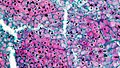
 Preadolescent Parathyroid Gland (32594358967).jpg 3,264 × 1,840; 5.4 MB
-
 Protective Coleoptile Tip in Zea Mays Embryo (40725880323).jpg 3,264 × 1,840; 906 KB
-
 Protective Coleoptile Tip in Zea Mays Embryo (47691344541).jpg 3,264 × 1,840; 1.17 MB
-
 Radicle or Embryonic Root in Zea Mays Embryo (47636870242).jpg 3,264 × 1,840; 1.37 MB
-
 Radicle With Inner Root Cap and Outer Coleorhiza in Zea Mays Embryo (32746619877).jpg 3,264 × 1,840; 5.4 MB
-
 Root parenchyma (34877668995).jpg 3,264 × 1,840; 716 KB
-
 Sclereids in Pyrus pear (36452689110).jpg 3,264 × 1,840; 4.19 MB
-
 Starch Granules in Endosperm of Zea Mays Embryo by Phase Contrast (40725881153).jpg 3,264 × 1,840; 1.78 MB
-
 Stele in Herbaceous Dicot root Ranunculus (34758989431).jpg 3,264 × 1,840; 1.06 MB
-
 Stomata and guard cells in upper epidermis of succulent (34810756046).jpg 3,264 × 1,840; 1.19 MB
-
 Stone cells in Pyrus pear (36452689990).jpg 3,264 × 1,840; 4.41 MB
-
 Tannin Cells in Sparganium (37403467520).jpg 3,264 × 1,840; 1.04 MB
-
 Thickened Bran in Kernel Stalk of Zea Mays Embryo (47639231542).jpg 3,264 × 1,840; 1.03 MB
-
 Typical Dicot Root Stele in Young Ranunculus (36006230295).jpg 3,264 × 1,840; 5.47 MB
-
 Typical Dicot Root Young Rananculus (36006227455).jpg 3,264 × 1,840; 5.34 MB
-
 Typical Dicot Root Young Ranunculus (36006229105).jpg 3,264 × 1,840; 1.93 MB
-
 Vascular Bundles in Zea (34837901016).jpg 3,264 × 1,840; 3.26 MB
-
 Vascular Strand in Cotyledon of Zea Mays Embryo (40724989953).jpg 3,264 × 1,840; 1.45 MB
-
 Vascular Strand in the Cotyledon of Zea Mays Embryo (46773820635).jpg 3,264 × 1,840; 4.35 MB
-
 Woody Dicot Root Cork in Quercus (36248758395).jpg 3,264 × 1,840; 1.21 MB
-
 Woody Dicot Root Medullary Rays in Quercus (36248757835).jpg 3,264 × 1,840; 1.29 MB
-
 Woody Dicot Root Quercus (36248754875).jpg 3,264 × 1,840; 7.51 MB
-
 Woody Dicot Root Quercus (36248756335).jpg 3,264 × 1,840; 2.02 MB
-
 Woody Dicot Root Quercus (36248759705).jpg 3,264 × 1,840; 1.22 MB
-
 Woody Dicot Root Vascular Cylinder in Quercus (36248755355).jpg 3,264 × 1,840; 1.88 MB
-
 Xylem vessels in developing Coleus shoot (35105390750).jpg 3,264 × 1,840; 2.04 MB
-
 Young Herbaceous Dicot Root Cortex in Ranunculus (34745655391).jpg 3,264 × 1,840; 5.48 MB
-
 Young Herbaceous Dicot Root Ranunculus (34745654101).jpg 3,264 × 1,840; 4.25 MB
-
 Young Herbaceous Dicot Root Ranunculus (34745654871).jpg 3,264 × 1,840; 6.63 MB
.jpg)
.jpg)
.jpg)
.jpg)
.jpg)
.jpg)
.jpg)
.jpg)
.jpg)
.jpg)
.jpg)
.jpg)
.jpg)
.jpg)
.jpg)
.jpg)
.jpg)
.jpg)
.jpg)
.jpg)
.jpg)
.jpg)
.jpg)
.jpg)
.jpg)
.jpg)
.jpg)
.jpg)
.png)
.jpg)
.jpg)
.jpg)
.jpg)
.jpg)
.jpg)
.jpg)
.jpg)
.jpg)
_(27847646938).jpg)
_(27847648428).jpg)
_(39909208420).jpg)
.jpg)
.jpg)
.jpg)
.jpg)
.jpg)
.jpg)
.jpg)
.jpg)
.jpg)
.jpg)
.jpg)
.jpg)
.jpg)
.jpg)
.jpg)
.jpg)
.jpg)
.jpg)
.jpg)
.jpg)
.jpg)
.jpg)
.jpg)
.jpg)
.jpg)
.jpg)
.jpg)
.jpg)
.jpg)
.jpg)
.jpg)
.jpg)
.jpg)
.jpg)
.jpg)
.jpg)
.jpg)
.jpg)
.jpg)
.jpg)
.jpg)
.jpg)
.jpg)
.jpg)
.jpg)
.jpg)
.jpg)
.jpg)
.jpg)
.jpg)
.jpg)
.jpg)
.jpg)
.jpg)
.jpg)
.jpg)
.jpg)
.jpg)
.jpg)
.jpg)
.jpg)
.jpg)
.jpg)
.jpg)
.jpg)
.jpg)
.jpg)
.jpg)
.jpg)
.jpg)
.jpg)
.jpg)
.jpg)
.jpg)
.jpg)
.jpg)
.jpg)
.jpg)
.jpg)
.jpg)
.jpg)
.jpg)
.jpg)
.jpg)
.jpg)
.jpg)
.jpg)
.jpg)
.jpg)
.jpg)
.jpg)
.jpg)
.jpg)
.jpg)
.jpg)
.jpg)
.jpg)
.jpg)
.jpg)
.jpg)
.jpg)
.jpg)
.jpg)
.jpg)
.jpg)
.jpg)
.jpg)
.jpg)
.jpg)
.jpg)
.jpg)
.jpg)
.jpg)
.jpg)
.jpg)
.jpg)
.jpg)
.jpg)
.jpg)
.jpg)
.jpg)
.jpg)
.jpg)