Category:Echinoidea anatomy
Jump to navigation
Jump to search
Subcategories
This category has the following 3 subcategories, out of 3 total.
A
- Aristotle's lantern (14 F)
E
Media in category "Echinoidea anatomy"
The following 64 files are in this category, out of 64 total.
-
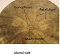
Aboraldendraster.jpg 405 × 375; 53 KB
-
AmbulacralfeetAmpulla.svg 350 × 288; 545 KB
-
Ambulacralsystem of Echinoidea.jpg 1,943 × 1,598; 1.05 MB
-
Arbacia lixula apical.JPG 578 × 439; 166 KB
-
Arbacia lixula valve.jpg 4,000 × 3,000; 3.16 MB
-
Arbacia punctulata 2.jpg 4,000 × 3,000; 4.12 MB
-
Archives de zoologie expérimentale et générale (1875) (20316741382).jpg 2,496 × 3,832; 1.85 MB
-
Brissus latecarinatus fasciole.jpg 2,668 × 2,000; 1.98 MB
-
Cake sand dollar (Arachnoides placenta) overturned and pecked.jpg 684 × 504; 134 KB
-
Dents d'oursin.jpg 1,200 × 919; 1.38 MB
-
Echinodermen (Stachelhäuter) (1889) (21146217521).jpg 1,974 × 3,170; 1.39 MB
-
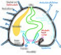
Echinoidea anatomie.svg 340 × 260; 19 KB
-
EchinoideaRegulariaCrossectionSchematik.svg 279 × 252; 112 KB
-
Echinus Mivart.png 916 × 640; 28 KB
-
Fish4572 - Flickr - NOAA Photo Library.jpg 684 × 539; 77 KB
-
Fish4573 - Flickr - NOAA Photo Library.jpg 649 × 537; 77 KB
-
Internal structuring of a Sea-biscuity thing.jpg 800 × 600; 77 KB
-
Lanterne d'Aristote de Toxopneustes pileolus.jpg 1,200 × 925; 1.01 MB
-
Lanterne d'Aristote.jpg 1,200 × 903; 764 KB
-
Mundfeld.jpg 853 × 684; 160 KB
-
Oraldendraster.jpg 523 × 370; 67 KB
-
Oursintortueinterneexterne.JPG 4,320 × 3,240; 2.66 MB
-
Paracentrotus lividus spines.JPG 1,895 × 1,502; 1.36 MB
-
Pedicellariae Mivart.png 302 × 1,310; 12 KB
-
Podia de Colobocentrotus atratus.JPG 1,200 × 900; 191 KB
-
Podia de Toxopneustes pileolus.jpg 1,200 × 918; 1.11 MB
-
PSM V13 D339 Sea urchin.jpg 1,466 × 583; 166 KB
-
PSM V13 D341 Cake and key hole urchin.jpg 1,713 × 893; 255 KB
-
PSM V27 D376 Pedicellariae.jpg 703 × 865; 46 KB
-
PSM V27 D378 Terminal portion of a tube foot.jpg 372 × 784; 69 KB
-
PSM V27 D379 An echinus partly denuded of its spines.jpg 1,045 × 953; 227 KB
-
PSM V27 D380 Sea urchin echinus.jpg 1,204 × 635; 105 KB
-
PSM V27 D381 Echinus raises itself.jpg 1,106 × 977; 187 KB
-
PSM V27 D382 Sea urchin curling up for protection.jpg 1,198 × 716; 146 KB
-
PSM V27 D383 Righting and ambulacral movements of segments of echinus.jpg 1,264 × 1,810; 275 KB
-
Pédicellaires (Haeckel).png 750 × 745; 706 KB
-
Pédicellaires de Toxopneustes pileolus.JPG 1,200 × 858; 882 KB
-
Radiole de Prionocidaris 1 (Haeckel).png 1,864 × 318; 472 KB
-
Radiole de Prionocidaris 2 (Haeckel).png 1,662 × 352; 568 KB
-
Radioles d'Heterocentrotus mamillatus sculptées.JPG 4,000 × 3,000; 2.74 MB
-
Radioles d'Heterocentrotus mamillatus.JPG 4,000 × 3,000; 3.3 MB
-
Sea Urchin Anatomy.svg 512 × 281; 131 KB
-
Sea urchin shell - pattern (6658690371).jpg 1,482 × 890; 246 KB
-
Sea Urchin Shell detail.jpg 5,184 × 3,456; 9.02 MB
-
Sea urchin.jpg 2,816 × 2,112; 2.77 MB
-
Sea-Urchin Case.JPG 2,448 × 3,264; 922 KB
-
Seeigel Gebiss.jpg 3,000 × 1,686; 3.34 MB
-
Seeigel-Bau.jpg 1,548 × 1,400; 884 KB
-
Seeigel-Saugfuesse(Galicien2005).jpg 616 × 616; 80 KB
-
Strongylocentrotus purpuratus 020313.JPG 2,028 × 1,566; 2.06 MB
-
Unknown Sand dollar 1 Macro.JPG 2,272 × 1,704; 1.23 MB
-
Urchin9b.jpg 900 × 606; 242 KB


