Category:Collections of Musée Dupuytren
Jump to navigation
Jump to search
Media in category "Collections of Musée Dupuytren"
The following 46 files are in this category, out of 46 total.
-
 Balles prélevées sur un soldat 36.jpg 3,000 × 2,003; 2.49 MB
-
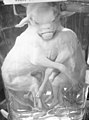
 Buste d’un homme probablement atteint de la variole 1.jpg 5,000 × 3,338; 5.53 MB
-
 Buste d’un homme probablement atteint de la variole 2.jpg 4,000 × 3,199; 3.81 MB
-
 Calcul volumineux du vagin avec fistule vésico-vaginale.jpg 5,000 × 3,347; 5.6 MB
-
 Cancer colloïde de l’épiploon et de l’estomac.jpg 4,000 × 4,000; 4.23 MB
-
 Cerveau de Louis Victor Leborgne dit Tantan 1.jpg 3,800 × 4,750; 4.64 MB
-
 Cerveau de Louis Victor Leborgne dit Tantan 2.jpg 4,500 × 3,600; 5.55 MB
-
 Cerveau de Louis Victor Leborgne dit Tantan 3.jpg 3,200 × 4,000; 3.99 MB
-
 Cerveau M. Leborgne.jpg 1,536 × 2,048; 790 KB
-
 Chaton à deux têtes.jpg 3,337 × 5,000; 4.52 MB
-
 Coeur présentant un rétrécissement auriculo-ventriculaire.jpg 3,600 × 4,500; 3.74 MB
-
 CollectionDupuytren 18.jpg 5,000 × 3,337; 4.37 MB
-
 CollectionDupuytren 35.jpg 5,000 × 3,337; 5.84 MB
-
 CollectionDupuytren 37.jpg 5,000 × 3,337; 6.4 MB
-
 CollectionDupuytren 38.jpg 5,000 × 3,337; 6.9 MB
-
 CollectionDupuytren 39.jpg 5,000 × 3,337; 5.83 MB
-
 Coupe d’un cerveau présentant une hémorragie cérébrale avec inondation ventriculaire.jpg 3,980 × 5,000; 5.21 MB
-
 Crâne présentant des nécroses ou caries syphilitiques 1.jpg 4,000 × 2,670; 2.26 MB
-
 Crâne présentant des nécroses ou caries syphilitiques 2.jpg 4,000 × 2,669; 2.07 MB
-
 Extrémité supérieure d’un fémur gauche 1.jpg 5,977 × 4,011; 6.72 MB
-
 Extrémité supérieure d’un fémur gauche 2.jpg 3,462 × 4,328; 4.17 MB
-
 Human conjoined twins DSC09364.jpg 719 × 1,203; 156 KB
-
 Humérus gauche présentant une nécrose 1.jpg 3,338 × 5,000; 4.91 MB
-
 Humérus gauche présentant une nécrose 2.jpg 3,440 × 5,153; 5.17 MB
-
 Kystes volumineux retirés d’un ovaire.jpg 5,000 × 3,338; 5.59 MB
-
 Lame histologique du cas Joffrin.jpg 4,635 × 3,094; 3.26 MB
-
 Lithiase rénale avec dilatation du bassinet et des calices.jpg 4,000 × 5,000; 5.39 MB
-
 Main droite atteinte de la lèpre 1.jpg 5,000 × 4,000; 5.72 MB
-
 Main droite atteinte de la lèpre 2.jpg 5,000 × 2,812; 3.09 MB
-
 Main gauche atteinte de radiodermite chronique hyperplasique.jpg 3,337 × 5,000; 4.67 MB
-
 MD.S.2015.0.2657 (1).jpg 3,904 × 3,360; 1.25 MB
-
 MD.S.2015.0.2787 (1).jpg 2,472 × 3,560; 726 KB
-
 Microtome de Minot.jpg 4,803 × 3,841; 5.45 MB
-
 Pipine Musée Dupuytren 2013.jpg 1,536 × 2,048; 656 KB
-
 Plaque photographique gélatino-bromure du cas Tissot.jpg 4,396 × 2,935; 2.83 MB
-
 Portion de colonne vertébrale atteinte du mal de Pott.jpg 4,703 × 4,702; 5.85 MB
-
 Portion de colonne vertébrale, 1863.jpg 3,549 × 4,437; 4.69 MB
-
 Poumon atteint de silicose.jpg 3,337 × 5,000; 5.15 MB
-
 Poumon atteint de tuberculose.jpg 4,000 × 5,000; 5.69 MB
-
 Quarante-cinq globes oculaires représentant une variété de pathologies.jpg 5,000 × 3,338; 5.36 MB
-
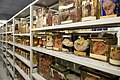 Réserve des préparations humides du Musée Dupuytren 1.jpg 5,000 × 3,337; 6.49 MB
-
 Réserve des préparations humides du Musée Dupuytren 2.jpg 5,000 × 3,337; 6.13 MB
-
 Siamese cattle twins DSC09327.jpg 904 × 1,214; 188 KB
-
 SU.MD.S.2015.0.2657 (1).jpg 4,188 × 3,384; 1.23 MB
-
 Tête d'un jeune sujet atteint d'une tumeur maxillaire 1.jpg 5,000 × 4,000; 5.44 MB
-
 Tête d'un jeune sujet atteint d'une tumeur maxillaire 2.jpg 3,601 × 4,500; 5.16 MB


.jpg)
.jpg)


.jpg)

