Category:Cloaca (embryo)
Jump to navigation
Jump to search
Subcategories
This category has the following 2 subcategories, out of 2 total.
G
- Genital tubercle (17 F)
U
- Urogenital sinus (43 F)
Media in category "Cloaca (embryo)"
The following 79 files are in this category, out of 79 total.
-
 A genetic fate map of Six2-expressing PCM progenitors.png 3,203 × 1,888; 2.6 MB
-
 A laboratory manual for comparative vertebrate anatomy (1922) (20754316592).jpg 1,408 × 414; 103 KB
-

-
-

-

-
 Activity of the BmpSmad and JNK pathways in the murine cloacal region.jpg 1,149 × 575; 530 KB
-
 An inducible genetic fate map of Six2-expressing PCM progenitors.png 1,793 × 1,379; 3.4 MB
-
 Bmp7 functions to promote cell survival in the cloacal endoderm.jpg 1,103 × 297; 321 KB
-

-
 Comparative anatomy (1936) (20661193782).jpg 1,742 × 408; 202 KB
-
 Comparative anatomy of vertebrates (1912) (20481863468).jpg 1,726 × 1,002; 283 KB
-
 Cunningham's Text-book of anatomy (1914) (20629759258).jpg 744 × 1,892; 222 KB
-
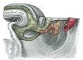
 Development of the Alimentary Canal, as seen in a Human Embryo about Five Weeks Old.jpg 1,274 × 1,012; 1.5 MB
-
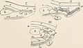
 Development of the cloaca different species.png 1,026 × 687; 361 KB
-

-

-

-

-

-

-
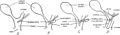
 Drei Schemata der Entwickelung der Urogenitalorgane.jpg 1,347 × 575; 689 KB
-

-

-
 Dynamic expression patterns of Six1 and Six2 during urogenital development.png 3,072 × 3,498; 3.61 MB
-
 Epithelial differentiation is delayed in Bmp7 null endoderm.jpg 1,010 × 801; 666 KB
-

-
 Gray1110.png 1,350 × 3,500; 1.24 MB
-
 Gray1115.png 888 × 672; 595 KB
-
 Gray1116.png 980 × 722; 1.02 MB
-
 Gray977.png 779 × 872; 540 KB
-
 Gray991.png 781 × 691; 609 KB
-
 Gray992.png 814 × 831; 587 KB
-

-
 Hypoplastic genital tubercles of Six1;Six2 compound mutants at e11.5.png 2,016 × 1,044; 4.19 MB
-
 Image from page 337 of "Chordate morphology" (1962) (19989513674).jpg 1,870 × 794; 344 KB
-
 Image from page 341 of "Chordate morphology" (1962) (20585824866).jpg 1,018 × 1,082; 746 KB
-

-
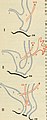
 Indifferentes Stadium der Urogenitalanlage nach Hertwig.png 575 × 787; 420 KB
-
 Longitudinal-section diagrams of the caudal half of the chick embryo.jpg 963 × 681; 386 KB
-

-

-
 Model for cloacal septation.png 2,080 × 1,414; 996 KB
-
 Polarity of cell divisions is disrupted in Bmp7 null the cloacal endoderm.jpg 1,135 × 1,473; 1.05 MB
-
 Sagittal Section of Human Zygote.jpg 817 × 684; 653 KB
-

-

-
 Schema of a Longitudinal Section of a Human Embryo.jpg 1,125 × 694; 1.02 MB
-

-

-

-

-
Sequential serial sections of ShhCreERT2+; R26LacZ+ embryos at E11.5.ogv 6.7 s, 640 × 370; 1.51 MB
-
 Six1 and Six2 are required for proliferation of PCM progenitors.png 2,569 × 1,001; 3.07 MB
-
 Six1;Six2 compound mutant genital tubercles have aberrant apoptosis patterns.png 3,582 × 2,055; 1.8 MB
-

-
 Tail End of a Human- Embryo about 25 Days Old.png 784 × 715; 872 KB
-
 Tail End of a Human-Embryo about 25 Days Old.jpg 769 × 702; 511 KB
-
 Tail End of Human Embryo about 33 Days Old.png 857 × 677; 754 KB
-

-

-
 Tail End op Female Human Embryo about 9 Weeks Old.png 1,259 × 763; 1.16 MB
-
 The cloacal morphology and connection sites of the nephric ducts.jpg 1,575 × 638; 420 KB
-
 The development of the chick; an introduction to embryology (1919) (14752383451).jpg 1,290 × 2,688; 349 KB
-

-

-
-
-
-
The three-dimensionally reconstructed cloaca at E10.5 Mouse.ogv 6.6 s, 640 × 444; 812 KB
-
The three-dimensionally reconstructed cloaca at E11.5 Mouse.ogv 6.5 s, 640 × 444; 677 KB
-
The three-dimensionally reconstructed cloaca at E9.5 Mouse.ogv 6.7 s, 640 × 444; 1.12 MB
-
The three-dimensionally reconstructed cloaca of ShhCreERT2flox, R26LacZ+ mutants at E13.5.ogv 6.7 s, 640 × 444; 1.26 MB
-
 Three-dimensional analyses of cloacal division processes.jpg 1,575 × 1,368; 1.35 MB
-

-
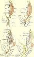
 Urogenitalanlage Entwickelung (01).jpg 1,249 × 795; 971 KB
-
 Urogenitalanlage Entwickelung.jpg 1,098 × 764; 730 KB
-

-


_(1907)_(20164950350).jpg)


_(20481863468).jpg)


_(20947658035).jpg)


_(19989513674).jpg)
_(20585824866).jpg)
_(20047305684).jpg)


_(14752383451).jpg)
_(20898803691).jpg)
_(14581562657).jpg)

,_and_arrest_in_cloacal_septation_in_Bmp7_null_embryo_(C_compare_to_B).jpg)
.jpg)


_(20754316592).jpg)
_(20661193782).jpg)
_(20629759258).jpg)