Category:Branchiostoma anatomy
Jump to navigation
Jump to search
Subcategories
This category has only the following subcategory.
Media in category "Branchiostoma anatomy"
The following 55 files are in this category, out of 55 total.
-
 1911 Britannica - Amphioxus lauceolatus.png 1,067 × 1,991; 285 KB
-
 1911 Britannica - Diagram of embryo of Amphioxus.png 516 × 1,089; 82 KB
-
 1911 Britannica - Pelagic larvae of A. lanceolatus.png 1,049 × 667; 63 KB
-
 1911 Britannica - Transverse sections of Amphioxus.png 1,086 × 1,891; 217 KB
-
 Amphioxus - Pharyngeal Region c.s..jpg 720 × 960; 86 KB
-
 Amphioxus bottom pharyngeal CS L.jpg 1,200 × 886; 143 KB
-
 Amphioxus bottom pharyngeal CS.jpg 1,200 × 886; 139 KB
-
 Amphioxus bottom pharyngeal zoom CS L.jpg 1,200 × 886; 194 KB
-
 Amphioxus bottom pharyngeal zoom CS.jpg 1,200 × 886; 193 KB
-
 Amphioxus cs Pharyngeal.jpg 1,179 × 1,528; 239 KB
-
 Amphioxus male CS L.jpg 787 × 1,494; 164 KB
-
 Amphioxus male CS.jpg 787 × 1,494; 164 KB
-
 Amphioxus pharyngeal CS L.jpg 1,027 × 1,200; 115 KB
-
 Amphioxus pharyngeal CS.jpg 1,027 × 1,200; 113 KB
-
 Amphioxus pharyngeal tail CS.jpg 1,026 × 1,200; 98 KB
-
 Amphioxus pharyngeal top CS L.jpg 1,200 × 886; 137 KB
-
 Amphioxus pharyngeal top CS.jpg 1,200 × 886; 133 KB
-
 Amphioxus pharyngeal top zoom CS L.jpg 1,200 × 886; 171 KB
-
 Amphioxus pharyngeal top zoom CS.jpg 1,200 × 886; 169 KB
-
 Amphioxus pharyngeal.jpg 1,200 × 886; 112 KB
-
 Amphioxus tail L.jpg 1,200 × 886; 70 KB
-
 Amphioxus tail.jpg 1,200 × 886; 67 KB
-
 Amphioxus whole L.jpg 1,200 × 628; 43 KB
-
 Amphioxus whole.jpg 1,200 × 628; 33 KB
-
 Animal biology (1938) (17574048064).jpg 3,200 × 1,278; 668 KB
-
 Anterior External L.jpg 1,075 × 1,445; 326 KB
-
 Branchiostoma cultellus1.jpg 1,000 × 768; 252 KB
-
 Branchiostoma lanceolatum (Pallas, 1774) - coupes transversales légendées.jpg 2,520 × 1,876; 3.86 MB
-
 Branchiostoma lanceolatum (Pallas, 1774) - coupes transversales.jpg 2,520 × 1,876; 3.73 MB
-
 Branchiostoma lanceolatum (Pallas, 1774) 1 - légendée.jpg 4,288 × 2,247; 3.17 MB
-
 Branchiostoma lanceolatum (Pallas, 1774) 3.jpg 3,620 × 1,344; 2.54 MB
-
 BranchiostomaLanceolatum PioM (hy).png 2,165 × 704; 286 KB
-
 Cepl003b.png 1,411 × 1,662; 182 KB
-
 Cepl013c.png 2,330 × 937; 484 KB
-
 Chor Cepl amphioxus head.jpg 2,048 × 1,536; 397 KB
-
 Cross section of Amphioxus.jpg 800 × 598; 74 KB
-
 Gonadal Section L.jpg 1,666 × 1,220; 379 KB
-
 Lancelet GFP GIF.gif 600 × 600; 4.07 MB
-
 Lancelet's endostyle1, numbered.jpg 950 × 1,200; 402 KB
-
 Lancelet's endostyle1.jpg 950 × 1,200; 444 KB
-
 Lancelet's endostyle2, numbered.jpg 950 × 1,200; 512 KB
-
 Lancelet's endostyle2.jpg 950 × 1,200; 583 KB
-
 Lancelet's gill slits, numbered.jpg 1,200 × 950; 455 KB
-
 Lancelet's gill slits.jpg 1,200 × 950; 508 KB
-
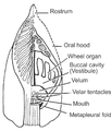
 Lancelet's metapleural fold.jpg 1,200 × 950; 356 KB
-
 Lancelet's mid-gut diverticulum.jpg 950 × 1,600; 801 KB
-
 Lancelet's nerve cord.jpg 1,200 × 950; 620 KB
-
 Lancelet's ovary.jpg 950 × 1,200; 383 KB
-
 Lectures on the elements of comparative anatomy (Page 190) BHL3318940.jpg 2,316 × 3,759; 738 KB
-
 Meyers b5 s0350 b1.png 570 × 218; 53 KB
-
 Tail Section CS L.jpg 1,491 × 1,198; 189 KB
-
 The Amphioxus and its development (1893) (17542538974).jpg 1,846 × 2,740; 1.2 MB
-
 The Amphioxus and its development (1893) (17544525153).jpg 1,776 × 2,738; 1.39 MB
-
 The Amphioxus and its development (1893) (17978889339).jpg 2,624 × 1,710; 1.2 MB
-
 Transverse section of lancelet.jpg 950 × 1,200; 287 KB


_1_-_l%C3%A9gend%C3%A9e.jpg)


_BHL3318940.jpg)

_(17542538974).jpg)
_(17544525153).jpg)
_(17978889339).jpg)
_(17574048064).jpg)
_-_coupes_transversales_l%C3%A9gend%C3%A9es.jpg)
_-_coupes_transversales.jpg)
_3.jpg)
.png)