Category:Black and white microscopic images
Jump to navigation
Jump to search
These are black and white micrographs.
Media in category "Black and white microscopic images"
The following 34 files are in this category, out of 34 total.
-
 BabbitB83.jpg 864 × 697; 229 KB
-
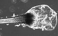
 Broken carbon fiber bar.jpg 435 × 383; 60 KB
-
 ExposureTimeSeries.tif 1,000 × 210; 225 KB
-
 Flattened cochlear preparation.png 855 × 1,360; 615 KB
-
 Fucus conceptacle.tif 900 × 900; 794 KB
-
 Hair Cell Patterning Defects in the Cochlea.png 2,012 × 2,475; 3.05 MB
-
 Hundehaar-Katzenhaar.jpg 600 × 484; 32 KB
-
 Inner ear pathology in MPS IIIB mice at 30 wks-A.png 1,985 × 1,112; 1.18 MB
-
 Inner ear pathology in MPS IIIB mice at 30 wks-B.png 1,986 × 1,324; 1.62 MB
-
 Inner ear pathology in MPS IIIB mice at 30 wks-C.png 1,970 × 1,416; 1.4 MB
-
 Inner ear pathology in MPS IIIB mice at 30 wks-D.png 1,986 × 1,516; 1.82 MB
-
 Inner ear pathology in MPS IIIB mice at 30 wks-E.png 1,969 × 2,421; 2.59 MB
-
 Inner ear pathology in MPS IIIB mice at 30 wks-F.png 1,986 × 777; 795 KB
-
 Inner ear pathology in MPS IIIB mice at 30 wks.png 3,971 × 4,961; 14.57 MB
-
 Katzenhaare unter dem Mikroskop.jpg 800 × 659; 46 KB
-
 Lokalisation der so charakteristischen Spirochaeten in Reinkultur.JPG 659 × 655; 267 KB
-
 Mammillaria sp., semeno (010).jpg 3,072 × 2,304; 4.54 MB
-
 Microcystis aeruginosa 680.jpg 1,024 × 768; 189 KB
-
 Microgroove.tif 512 × 480; 263 KB
-
 Microstructure steel annealed nut.jpg 1,700 × 2,856; 3.77 MB
-
 Nähzwirn 3-fach.jpeg 1,259 × 815; 96 KB
-
 Piqué Mikroskopaufnahme.jpeg 1,251 × 818; 104 KB
-
 PSM V05 D666 Melicerta ringens.jpg 1,675 × 3,458; 628 KB
-
 PSM V05 D668 Flosculaeia ornata.jpg 2,077 × 3,464; 640 KB
-
 PSM V35 D267 Amoebae of well mud.jpg 374 × 642; 26 KB
-
 PSM V35 D268 Centropyxis aculeata and flagellate infusoria.jpg 1,708 × 599; 89 KB
-
 PSM V35 D268 Shell bearing amoeba euglypha.jpg 564 × 447; 29 KB
-
 PSM V35 D269 Cothurnia and stenostonia leucops.jpg 1,062 × 965; 84 KB
-
 PSM V35 D269 Crab like cyclops.jpg 990 × 1,243; 95 KB
-
 PSM V35 D270 Crab like cypris.jpg 985 × 494; 72 KB
-
 Tierhaar unter dem Mikroskop.jpg 800 × 501; 62 KB
-
 Troostit.jpg 960 × 768; 403 KB
-
 Viscosa2.JPG 391 × 284; 16 KB
-
 Widmannstaetten.png 1,024 × 819; 784 KB


.jpg)


