Category:Biological illustrations by DrJanaOfficial
Jump to navigation
Jump to search
Media in category "Biological illustrations by DrJanaOfficial"
The following 102 files are in this category, out of 102 total.
-
 3d Scapula Rendered.gif 614 × 614; 4.81 MB
-
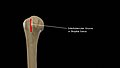
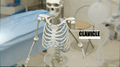
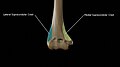
 Acromion End of the Clavicle.png 1,920 × 1,080; 2.16 MB
-
 Alzheimer's Disease.gif 1,024 × 778; 4.59 MB
-
 Anatomy of Scapula.gif 600 × 338; 1.74 MB
-
 Animated Heart - Old Textbook style.gif 2,560 × 1,866; 6.2 MB
-
 Anterior View of Right Distal Radius.png 1,152 × 2,048; 1.63 MB
-
 Anterior View of Right Proximal Radius.png 1,152 × 2,048; 1.36 MB
-
 Anterior-view-of-the-humerus-showing-borders-and-surfaces.jpg 1,743 × 937; 443 KB
-
 Atlas The First Cervical Vertebra.gif 800 × 800; 7.15 MB
-
 Attachments-of-greater-tubercle.jpg 1,787 × 853; 454 KB
-
 Bicipital-groove.jpg 1,570 × 892; 336 KB
-
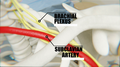 Brachial Plexus.png 1,920 × 1,080; 2.02 MB
-
 Capitulum-and-trochlea.jpg 1,554 × 873; 323 KB
-
 Cardiac-Cycle-Animated.gif 1,024 × 1,536; 6.11 MB
-
 Cervica lVertebrae.jpg 1,600 × 1,200; 323 KB
-
 Cervical Spine - Atlas and Axis.png 4,096 × 4,096; 31.76 MB
-
 Cervical Spine -C3 and C4 vertebrae along with uncinate process.png 4,096 × 4,096; 31.1 MB
-
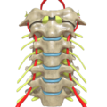 Cervical Spine Anterior View.png 4,096 × 4,096; 18.14 MB
-
 Cervical Spine Bottom View.png 4,096 × 4,096; 17.42 MB
-
 Cervical Spine Computer Generated Image.png 2,048 × 2,048; 2.79 MB
-
 Cervical Spine Cross View.png 4,096 × 4,096; 29.35 MB
-
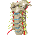
 Cervical Spine Lateral View.png 4,096 × 4,096; 10.3 MB
-
 Cervical Spine Oblique View.png 4,096 × 4,096; 18.65 MB
-
 Cervical Spine Side View.png 4,096 × 4,096; 8.87 MB
-
 Cervical Vertebrae C5 and C6.png 4,096 × 4,096; 28.79 MB
-
 Cervical Vertebrae.png 4,096 × 4,096; 31.63 MB
-
 CervicalVertebrae.jpg 2,400 × 1,800; 897 KB
-
 CervicalVertebrae2.jpg 1,600 × 1,200; 247 KB
-
 CervicalVertebrae4.jpg 1,600 × 1,200; 456 KB
-
 CervicalVertebrae5.jpg 1,600 × 1,200; 478 KB
-
 Clavicle 3d Model.gif 740 × 460; 10.19 MB
-
 Clavicle Highlighted.png 1,920 × 1,080; 1.86 MB
-
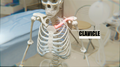
 Clavicle.png 1,920 × 1,080; 1.9 MB
-
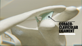 Coraco Clavicular Ligament.png 1,920 × 1,080; 1.98 MB
-
 Coronoid-Fossa.jpg 1,561 × 838; 336 KB
-
 Crest-for-lateral-head-of-triceps-brachii.jpg 1,394 × 867; 320 KB
-
 Crest-of-greater-tubercle-and-lesser-tubercle.jpg 1,664 × 937; 360 KB
-
 Crests-of-humerus.jpg 1,664 × 937; 405 KB
-
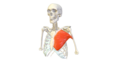
 Deltoid Muscle.png 1,920 × 1,080; 1.74 MB
-
 Deltoid-Tuberosity-Insertion-of-deltoideus-muscle.jpg 1,216 × 913; 266 KB
-
 Deltoid-Tuberosity.jpg 1,216 × 913; 263 KB
-
 DeltoPectoralTriangle.png 1,920 × 1,080; 2.29 MB
-
 Full Anterior View of Right Radius.png 1,152 × 2,048; 477 KB
-
 Full Lateral View of Right Radius.png 1,152 × 2,048; 402 KB
-
 Full Medial View of Right Radius.png 1,152 × 2,048; 420 KB
-
 Full Posterior View of Right Radius.png 1,152 × 2,048; 441 KB
-
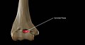
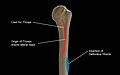
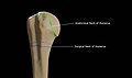
 Greater-Tubercle-of-Right-Humerus.jpg 1,575 × 840; 40 KB
-
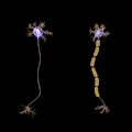
 Guillain-barré syndrome - Nerve Damage.gif 500 × 282; 3.42 MB
-
 Humerus 3d Model.gif 230 × 920; 6.29 MB
-
 Humerus-Head.gif 614 × 614; 8.84 MB
-
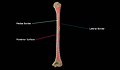
 Humerus-views.png 1,476 × 921; 873 KB
-
 Insertion-of-subscapularis-muscle.jpg 1,390 × 823; 289 KB
-
 Lateral View of Right Distal Radius.png 1,152 × 2,048; 923 KB
-
 Lateral View of Right Proximal Radius.png 1,152 × 2,048; 892 KB
-
 Lesser-Tubercle-of-Right-Humerus.jpg 1,390 × 823; 278 KB
-
 Medial View of Right Distal Radius.png 1,152 × 2,048; 1.34 MB
-
 Medial View of Right Proximal Radius.png 1,152 × 2,048; 1.46 MB
-
 Medial-epicondyle-and-lateral-epicondyle.jpg 1,554 × 873; 331 KB
-
 Medial-supracondylar-crest-and-lateral-supracondylar-crest.jpg 1,554 × 873; 342 KB
-
 Neck-of-Humerus.jpg 1,550 × 915; 48 KB
-
 Neuro Muscular Junction - Not Labelled.jpg 1,024 × 778; 164 KB
-
 Neuro Muscular Junction.png 4,096 × 3,112; 10.18 MB
-
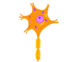 Neuron - Nerve Cell 01.png 4,096 × 3,112; 387 KB
-
 Neuron - Nerve Cell 02.png 2,048 × 1,556; 183 KB
-
 Neuron - Nerve Cell 03.png 1,024 × 778; 81 KB
-
 Neuron - Nerve Cell 04.png 512 × 389; 36 KB
-
 Neuron - Nerve Cell 05.png 256 × 195; 16 KB
-
 Neuron or Nerve cell - Labelled Image.png 4,096 × 3,112; 501 KB
-
 Nutrient-Foramen.jpg 1,457 × 877; 292 KB
-
 Olecranon-fossa.jpg 1,596 × 851; 338 KB
-
 Parts Of Clavicle.png 1,920 × 1,080; 2.32 MB
-
 Pectoralis Major Muscle.png 1,920 × 1,080; 1.87 MB
-
 Pectoralis Major.png 1,920 × 1,080; 735 KB
-
 PectoralisMajor.gif 640 × 360; 1.97 MB
-
 Posterior Radius Proximal.png 1,920 × 1,080; 271 KB
-
 Posterior View of Right Distal Radius.png 1,152 × 2,048; 1.04 MB
-
 Posterior View of Right Proximal Radius.png 1,152 × 2,048; 931 KB
-
 Posterior-view-of-the-humerus-showing-borders-and-surfaces.jpg 1,597 × 929; 376 KB
-
 Pronation and Supination.gif 800 × 450; 1.82 MB
-
 Radial-Fossa.jpg 1,584 × 871; 330 KB
-
 Radial-groove-continuing-as-lateral-border-of-shaft.jpg 1,438 × 837; 293 KB
-
 Radius and Ulna - Forearm Bones.jpg 2,048 × 2,048; 179 KB
-
 Radius Anteiror Distal 01.png 1,920 × 1,080; 294 KB
-
 Radius Anteiror Proximal.png 1,920 × 1,080; 276 KB
-
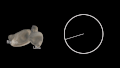 Radius Bone and Radius of a circle comparison.gif 800 × 450; 1.7 MB
-
 Radius Bone with Labels.gif 800 × 800; 2.85 MB
-
 Radius Posterior Distal.png 1,920 × 1,080; 334 KB
-
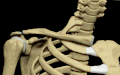
 Ribcage with both humerii.png 1,024 × 1,024; 1.34 MB
-
 Saltatory Conduction.gif 1,200 × 1,200; 1.83 MB
-
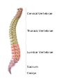 Segments of Vertebrae.svg 744 × 1,052; 281 KB
-
 Static Old Textbook Styled Human heart.jpg 1,280 × 933; 483 KB
-
 Sternocleidomastoid.png 1,920 × 1,080; 2.09 MB
-
 SternoHyoid.png 1,920 × 1,080; 1.98 MB
-
 Sternum.gif 800 × 800; 3.07 MB
-
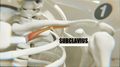 Subclavius.png 1,920 × 1,080; 2.12 MB
-
 Thoracic Cage with Both Humerii.gif 800 × 800; 7.84 MB
-
 Thoracic Cage with Spine - Anatomy.gif 800 × 800; 7.33 MB
-
 Trapezius Muscle Labelled.png 1,920 × 1,080; 2.13 MB
-
 Trapezius Muscle.png 1,920 × 1,080; 1.91 MB
-
 Trochlea.jpg 1,735 × 856; 429 KB
-
 Upper Limb Bones with articular cartilage.svg 3,200 × 3,200; 2.02 MB
-
 Upper Limb Bones.svg 3,200 × 3,200; 2.03 MB


