Category:Atomic force micrographs
Jump to navigation
Jump to search
Subcategories
This category has only the following subcategory.
A
- Atomic force microscopic videos (32 F)
Media in category "Atomic force micrographs"
The following 59 files are in this category, out of 59 total.
-

-
 24 02 06 10pc wk2 tap 002 - 1um x 170nm hap mx (214911319).jpg 888 × 888; 57 KB
-
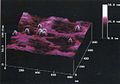
 3D AFM (30546875525).jpg 1,413 × 1,047; 69 KB
-
 3d severe deformed copper AFM.jpg 1,024 × 768; 58 KB
-
 3D Topografic AFM image.jpg 488 × 488; 31 KB
-
 A-typical-AFM-image-for-Ge-surface.jpg 600 × 709; 65 KB
-

-

-
 AFM 3D Topography Image of Palladium Nanoparticles on Chitosan Film.tif 1,024 × 718; 582 KB
-
 AFM 3D Topography Image of Silver Nanoparticles on Chitosan Film.tif 1,024 × 714; 440 KB
-
 Afm cd pits.jpg 256 × 256; 16 KB
-
 Afm cd-rom.jpg 2,655 × 2,113; 1.79 MB
-
 Afm compact-disc mrtz.png 1,030 × 815; 113 KB
-
 AFM image of a Bluray.jpg 612 × 514; 56 KB
-
 AFM image of a CCD-Chip.jpg 608 × 514; 37 KB
-
 AFM image of a DVD.jpg 608 × 514; 36 KB
-
 AFM image of tin layer.jpg 2,000 × 2,000; 526 KB
-
 AFM image ofwing butterfly.jpg 784 × 767; 263 KB
-
 AFM images of different magnetron sputtered lead layers.jpg 3,976 × 2,008; 1.48 MB
-
 AFM Innventia nanocellulose.JPG 419 × 421; 149 KB
-
 AFM MagneticMode HD.JPG 680 × 169; 18 KB
-

-
 AFM1.jpg 679 × 402; 54 KB
-
 AFM3.jpg 679 × 398; 46 KB
-
 AFMi 3D kujutis sadestatud üliõhukese polümeerkile servast.jpg 1,238 × 774; 368 KB
-
 AFMimageRoughGlass20x20.JPG 398 × 308; 41 KB
-
 AFMimageRoughGlass20x20.png 401 × 326; 187 KB
-
 AFMprzyklad.png 350 × 308; 65 KB
-
 Atomic Force Microscope (5940987164).jpg 620 × 308; 185 KB
-
 Atomic force microscope lithography.jpg 2,396 × 2,396; 869 KB
-
 Band 3 Atomic microscope.jpg 554 × 387; 32 KB
-
 Bilayer AFM schematic.png 2,391 × 1,503; 1.43 MB
-
 Bimodal AFM nanomechanical map of block-copolymer.png 308 × 599; 352 KB
-
 Bimodal AFM nanomechanical mapping of MoS2 on Si-SiO2.webp 1,910 × 645; 150 KB
-
 CD AFM.png 758 × 760; 351 KB
-
 Compact disk data layer 2d 3d.PNG 327 × 611; 166 KB
-
 DVD AFM J REBIS.png 1,121 × 746; 212 KB
-
 EUV mask blank - as polished.png 347 × 370; 216 KB
-
 EUV mask blank - untreated.png 449 × 434; 244 KB
-
 FromJAPANwithLOVE.jpg 2,379 × 2,379; 1.61 MB
-
 Hexabenzocoronene AFM 2.jpg 786 × 777; 100 KB
-
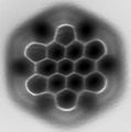 Hexabenzocoronene AFM.jpg 710 × 717; 77 KB
-
 Latex afm1.jpg 800 × 525; 73 KB
-
 MgAl2O3 AFM image.jpg 1,324 × 705; 45 KB
-
 MgO AFM image.jpeg 1,324 × 705; 61 KB
-
 Microscopy, Scanning Photoionization Imagery, SPIM Image (5940488089).jpg 1,452 × 828; 494 KB
-
 Mixed monolayer.jpg 3,600 × 3,600; 5.45 MB
-
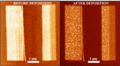 Mn12 before and after.png 832 × 460; 670 KB
-
 Nanobouillon (8537156410).jpg 3,900 × 3,900; 3.64 MB
-
 NanoScanHrMFMWD3200BEVT.JPG 512 × 512; 131 KB
-

-
 PI-number-LON.png 1,390 × 1,149; 1.2 MB
-
 Selenium nanosystem.png 525 × 256; 168 KB
-
 Silicon nanowire transistor in the shape of the word NANO.jpg 265 × 268; 13 KB
-
 Surface plexiglas sable AFM 3D.png 2,080 × 1,416; 1.19 MB
-
 Surface plexiglas sable AFM.png 256 × 256; 61 KB
-
 THz Antenne.jpg 764 × 568; 45 KB
-
 Two AFM images on 111 GaAs dots done at NIMs, Japan.png 271 × 824; 232 KB
-
 Активный тромбоцит.png 991 × 810; 1.4 MB
_xy_620nm_z_160nm_(214911321).jpg)
.jpg)
.jpg)


.jpg)


.jpg)


.jpg)

_xy_5um_z_2um_(214911322).jpg)


