Category:Anatomy of the human pelvic region
Jump to navigation
Jump to search
Subcategories
This category has the following 14 subcategories, out of 14 total.
A
B
C
H
J
M
P
V
X
Media in category "Anatomy of the human pelvic region"
The following 98 files are in this category, out of 98 total.
-
(155) Stylized depiction of action of puborectalis sling.png 1,692 × 1,484; 184 KB
-
A textbook of obstetrics (1898) (14780177172).jpg 2,004 × 2,012; 764 KB
-
Anatomie kostí pánevního kloubu.png 1,206 × 869; 718 KB
-
Bekkenledd.gif 300 × 262; 25 KB
-
Bourgery & Jacob-v10.jpg 2,823 × 2,413; 4.16 MB
-
Braus 1921 269.png 908 × 1,160; 3.02 MB
-
Building Fairs Brno 2011 (213).jpg 1,989 × 3,141; 3.42 MB
-
Buried penis on Xray KUB with multiple skin folds.jpg 3,456 × 4,608; 3.47 MB
-
Cephalic presentation.svg 263 × 232; 22 KB
-
Cephalicpre.JPG 342 × 598; 76 KB
-
Childbirth, from H. Deventer "Operationes Chirurgicae..." Wellcome L0013513.jpg 1,198 × 1,537; 687 KB
-
Clinical gyncology, medical and surgical (1895) (14784270172).jpg 1,582 × 2,320; 176 KB
-
Die Frau als Hausärztin (1911) 182 Tasterzirkel zur Beckenmessung.png 271 × 295; 123 KB
-
Dixon's Manual of human osteology (1912) - Fig 061.png 1,398 × 1,857; 1.53 MB
-
Dixon's Manual of human osteology (1912) - Fig 062.png 1,437 × 1,824; 1.04 MB
-
Female pelvic cavity.jpg 960 × 720; 116 KB
-
Female pelvis.png 780 × 1,000; 2.99 MB
-
Gerrish's Text-book of Anatomy (1902) - Fig. 182.png 1,244 × 1,132; 575 KB
-
Gerrish's Text-book of Anatomy (1902) - Fig. 183.png 1,444 × 1,044; 1.2 MB
-
Grant 1962 193.1.png 1,584 × 1,529; 1.18 MB
-
Grant 1962 194.png 4,147 × 3,223; 13.1 MB
-
Grant 1962 195.png 1,881 × 2,805; 2.54 MB
-
Grant 1962 196.1.png 1,749 × 1,738; 1.2 MB
-
Grant 1962 197.png 2,981 × 2,167; 3.44 MB
-
Grant 1962 198.png 3,421 × 2,101; 2.97 MB
-
Grant 1962 199.1.png 2,134 × 2,893; 6.38 MB
-
Grant 1962 200.png 3,619 × 2,079; 6.75 MB
-
Grant 1962 201.png 4,334 × 3,146; 13.21 MB
-
Grant 1962 202.png 4,378 × 3,432; 20.43 MB
-
Grant 1962 205.png 4,301 × 4,026; 24.52 MB
-
Grant 1962 206.png 1,474 × 2,552; 2.44 MB
-
Grant 1962 207.png 2,937 × 2,387; 8.56 MB
-
Grant 1962 208.1.png 1,628 × 1,617; 1.23 MB
-
Grant 1962 208.2.png 1,485 × 1,463; 770 KB
-
Grant 1962 210.png 3,971 × 3,069; 15 MB
-
Grant 1962 211.png 3,278 × 2,090; 2.6 MB
-
Grant 1962 212a.png 1,892 × 1,991; 1.9 MB
-
Grant 1962 212b.png 2,233 × 2,002; 2.51 MB
-
Grant 1962 213.png 4,472 × 4,632; 28.53 MB
-
Grant 1962 214-ar.png 2,651 × 3,168; 1.38 MB
-
Grant 1962 214.png 2,651 × 3,168; 1.48 MB
-
Grant 1962 215-ar.png 2,519 × 1,969; 1.29 MB
-
Grant 1962 215.png 2,519 × 1,969; 1.33 MB
-
Grant 1962 216.png 3,388 × 3,069; 13.68 MB
-
Grant 1962 217.png 3,520 × 2,662; 10.11 MB
-
Grant 1962 224.png 1,980 × 1,573; 2.96 MB
-
Grant 1962 225.png 4,466 × 3,740; 21.03 MB
-
Grant 1962 226.png 4,776 × 3,720; 20.41 MB
-
Grant 1962 227.png 4,376 × 3,888; 22.67 MB
-
Grant 1962 228.png 4,040 × 3,816; 14.56 MB
-
Grant 1962 229.png 4,536 × 4,016; 15.93 MB
-
Grant 1962 230.1.png 4,320 × 2,264; 13.02 MB
-
Grant 1962 231.png 4,884 × 4,422; 35.12 MB
-
Grant 1962 232.png 4,488 × 2,336; 6.86 MB
-
Grant 1962 233.png 4,560 × 3,264; 17.59 MB
-
Grant 1962 234.png 4,631 × 3,784; 24.61 MB
-
Grant 1962 235.png 4,587 × 4,532; 32.1 MB
-
Grant 1962 237.1.png 3,047 × 2,167; 6.75 MB
-
Grant 1962 238.png 4,554 × 3,784; 26.75 MB
-
Grant 1962 239.png 3,938 × 3,300; 13.17 MB
-
Grant 1962 240.png 3,388 × 3,190; 12.08 MB
-
Grant 1962 241.png 4,389 × 4,004; 18.09 MB
-
Gray408-ar.png 460 × 500; 133 KB
-
Gray408.png 460 × 500; 58 KB
-
Gynecological diagnosis (1910) (14778287545).jpg 2,024 × 1,452; 550 KB
-
Holden's human osteology (1899) - Fig27.png 500 × 1,046; 176 KB
-
Holden's human osteology (1899) - Fig28.png 249 × 1,152; 67 KB
-
Holden's human osteology (1899) - Fig29.png 642 × 909; 317 KB
-
Human coccyx - 4691.jpg 1,020 × 1,047; 1.46 MB
-
J.F. Gautier D'Agoty, Myologie complette en coleur... Wellcome L0023743.jpg 1,186 × 1,538; 636 KB
-
Male pelvic cavity.jpg 960 × 720; 124 KB
-
Merkel's Human Anatomy (1913) - Vol 3 - Fig 118.png 1,664 × 2,208; 675 KB
-
Operative gynecology - (1906) (14783482785).jpg 2,026 × 2,624; 1.1 MB
-
Pathology and treatment of diseases of women (1912) (14594959877).jpg 2,012 × 1,338; 536 KB
-
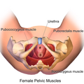
Pelvic Muscles (Female Inferior).png 1,500 × 1,500; 1.23 MB
-
Pelvic Muscles (Female Side) (cropped).png 1,500 × 1,355; 1.07 MB
-
Pelvic Muscles (Female Side).png 1,500 × 1,500; 1.15 MB
-
Pelvic Muscles (Male Side).png 1,500 × 1,500; 1.31 MB
-
Pelvis - os coxae, os sacrum (caudal view).jpg 4,608 × 3,456; 4.54 MB
-
Pelvis RG MR CT3d front 01.jpg 417 × 944; 44 KB
-
Pelvis; seven figures. Line engraving by Campbell, 1816-1821 Wellcome V0007949EL.jpg 1,184 × 1,640; 1.42 MB
-
Pelvix and Lower Spine of Human Embryo (3460140262).jpg 1,469 × 1,632; 1.19 MB
-
Small Lumbar Scoliosis Curve with Pelvic Torque.png 712 × 830; 396 KB
-
Sobo 1906 498.png 2,595 × 1,947; 4.83 MB
-
Sobo 1906 499.png 2,388 × 1,962; 4.48 MB
-
Sobo 1906 513.png 2,286 × 1,806; 3.95 MB
-
Sobo 1909 246.png 2,160 × 1,644; 10.18 MB
-
Sobo 1909 300.png 1,532 × 1,264; 5.55 MB
-
Sobo 1909 568.png 1,244 × 1,410; 5.03 MB
-
The bones of the pelvis. Engraving by G. Bartoli. Wellcome V0007880.jpg 3,200 × 2,190; 2.91 MB
-
The pelvis of an articulated skeleton. Drawing, ca. 1560 (?) Wellcome L0027180.jpg 2,978 × 3,886; 3.64 MB
-
The practice of surgery (1910) (14798809963).jpg 1,742 × 2,068; 647 KB
-
The bones of the pelvis. Engraving by G. Bartoli. Wellcome V0007881.jpg 3,213 × 2,175; 3.05 MB
-
Wiki directional muscle forcesfig2.jpg 1,721 × 1,258; 587 KB
-
Xray of pelvis with left sided phlebolith with some important lines.jpg 3,317 × 3,713; 5.94 MB


